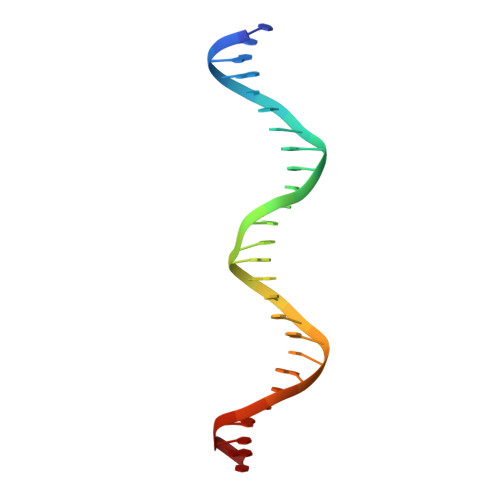
1H89

CRYSTAL STRUCTURE OF TERNARY PROTEIN-DNA COMPLEX2


Experimental Data Snapshot

  • Method: X-RAY DIFFRACTION
  • Resolution: 2.45 Å
  • R-Value Free: 0.267 
  • R-Value Work: 0.229 
  • R-Value Observed: 0.229 

wwPDB Validation   3D Report Full Report


This is version 1.4 of the entry. See complete history


Literature

Mechanism of C-Myb-C/Ebpbeta Cooperation from Separated Sites on a Promoter

Tahirov, T.H.Sato, K.Ichikawa-Iwata, E.Sasaki, M.Inoue-Bungo, T.Shiina, M.Kimura, K.Takata, S.Fujikawa, A.Morii, H.Kumasaka, T.Yamamoto, M.Ishii, S.Ogata, K.

(2002) Cell 108: 57

  • DOI: https://doi.org/10.1016/s0092-8674(01)00636-5
  • Primary Citation of Related Structures:  
    1H88, 1H89, 1H8A

  • PubMed Abstract: 

    c-Myb, but not avian myeloblastosis virus (AMV) v-Myb, cooperates with C/EBP beta to regulate transcription of myeloid-specific genes. To assess the structural basis for that difference, we determined the crystal structures of complexes comprised of the c-Myb or AMV v-Myb DNA-binding domain (DBD), the C/EBP beta DBD, and a promoter DNA fragment. Within the c-Myb complex, a DNA-bound C/EBP beta interacts with R2 of c-Myb bound to a different DNA fragment; point mutations in v-Myb R2 eliminate such interaction within the v-Myb complex. GST pull-down assays, luciferase trans-activation assays, and atomic force microscopy confirmed that the interaction of c-Myb and C/EBP beta observed in crystal mimics their long range interaction on the promoter, which is accompanied by intervening DNA looping.


  • Organizational Affiliation

    Kanagawa Academy of Science and Technology, 3-9 Fukuura, Kanazawa-ku, Yokohama 236-0004, Japan. tahir@med.yokohama-cu.ac.jp


Macromolecules

Find similar proteins by:  (by identity cutoff)  |  3D Structure
Entity ID: 1
MoleculeChains Sequence LengthOrganismDetailsImage
CAAT/ENHANCER BINDING PROTEIN BETA
A, B
64Homo sapiensMutation(s): 0 
UniProt & NIH Common Fund Data Resources
Find proteins for P17676 (Homo sapiens)
Explore P17676 
Go to UniProtKB:  P17676
PHAROS:  P17676
GTEx:  ENSG00000172216 
Entity Groups  
Sequence Clusters30% Identity50% Identity70% Identity90% Identity95% Identity100% Identity
UniProt GroupP17676
Sequence Annotations
Expand
  • Reference Sequence
Find similar proteins by:  (by identity cutoff)  |  3D Structure
Entity ID: 2
MoleculeChains Sequence LengthOrganismDetailsImage
MYB PROTO-ONCOGENE PROTEIN159Mus musculusMutation(s): 2 
UniProt & NIH Common Fund Data Resources
Find proteins for P06876 (Mus musculus)
Explore P06876 
Go to UniProtKB:  P06876
IMPC:  MGI:97249
Entity Groups  
Sequence Clusters30% Identity50% Identity70% Identity90% Identity95% Identity100% Identity
UniProt GroupP06876
Sequence Annotations
Expand
  • Reference Sequence
Find similar nucleic acids by:  (by identity cutoff)  |  3D Structure
Entity ID: 3
MoleculeChains LengthOrganismImage
DNA(5'-(*GP*AP*TP*GP*TP*GP*GP*CP*GP*CP*AP* AP*TP*CP*CP*TP*TP*AP*AP*CP*GP*GP*AP*CP*TP*G)-3')26Homo sapiens
Sequence Annotations
Expand
  • Reference Sequence
Find similar nucleic acids by:  (by identity cutoff)  |  3D Structure
Entity ID: 4
MoleculeChains LengthOrganismImage
DNA(5'-(*CP*CP*AP*GP*TP*CP*CP*GP*TP*TP*AP* AP*GP*GP*AP*TP*TP*GP*CP*GP*CP*CP*AP*CP*AP*T)-3')26Homo sapiens
Sequence Annotations
Expand
  • Reference Sequence
Small Molecules
Binding Affinity Annotations 
IDSourceBinding Affinity
DNA PDBBind:  1H89 Kd: 1.64 (nM) from 1 assay(s)
Experimental Data & Validation

Experimental Data

  • Method: X-RAY DIFFRACTION
  • Resolution: 2.45 Å
  • R-Value Free: 0.267 
  • R-Value Work: 0.229 
  • R-Value Observed: 0.229 
  • Space Group: P 21 21 21
Unit Cell:
Length ( Å )Angle ( ˚ )
a = 62.684α = 90
b = 73.383β = 90
c = 161.699γ = 90
Software Package:
Software NamePurpose
CNSrefinement
DENZOdata reduction
SCALEPACKdata scaling
X-PLORphasing

Structure Validation

View Full Validation Report


Entry History 

Deposition Data

Revision History  (Full details and data files)

  • Version 1.0: 2002-01-28
    Type: Initial release
  • Version 1.1: 2011-05-08
    Changes: Version format compliance
  • Version 1.2: 2011-07-13
    Changes: Version format compliance
  • Version 1.3: 2019-05-08
    Changes: Data collection, Experimental preparation, Other, Source and taxonomy
  • Version 1.4: 2023-12-13
    Changes: Data collection, Database references, Derived calculations, Other, Refinement description