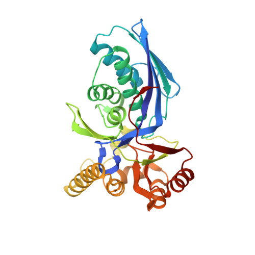
1H73

CRYSTAL STRUCTURE OF HOMOSERINE KINASE COMPLEXED WITH THREONINE


Experimental Data Snapshot

  • Method: X-RAY DIFFRACTION
  • Resolution: 2.00 Å
  • R-Value Free: 0.222 
  • R-Value Work: 0.178 
  • R-Value Observed: 0.178 

wwPDB Validation   3D Report Full Report


Ligand Structure Quality Assessment 


This is version 1.4 of the entry. See complete history


Literature

Structural Basis for the Catalysis and Substrate Specificity of Homoserine Kinase

Krishna, S.S.Zhou, T.Daugherty, M.Osterman, A.L.Zhang, H.

(2001) Biochemistry 40: 10810

  • DOI: https://doi.org/10.1021/bi010851z
  • Primary Citation of Related Structures:  
    1H72, 1H73, 1H74

  • PubMed Abstract: 

    Homoserine kinase (HSK), the fourth enzyme in the aspartate pathway of amino acid biosynthesis, catalyzes the phosphorylation of L-homoserine (Hse) to L-homoserine phosphate, an intermediate in the production of L-threonine, L-isoleucine, and in higher plants, L-methionine. The high-resolution structures of Methanococcus jannaschii HSK ternary complexes with its amino acid substrate and ATP analogues have been determined by X-ray crystallography. These structures reveal the structural determinants of the tight and highly specific binding of Hse, which is coupled with local conformational changes that enforce the sequestration of the substrate. The delta-hydroxyl group of bound Hse is only 3.4 A away from the gamma-phosphate of the bound nucleotide, poised for the in-line attack at the gamma-phosphorus. The bound nucleotides are flexible at the triphosphate tail. Nevertheless, a Mg(2+) was located in one of the complexes that binds between the beta- and gamma-phosphates of the nucleotide with good ligand geometry and is coordinated by the side chain of Glu130. No strong nucleophile (base) can be located near the phosphoryl acceptor hydroxyl group. Therefore, we propose that the catalytic mechanism of HSK does not involve a catalytic base for activating the phosphoryl acceptor hydroxyl but instead is mediated via a transition state stabilization mechanism.


  • Organizational Affiliation

    Department of Biochemistry, University of Texas Southwestern Medical Center, 5323 Harry Hines Boulevard, Dallas, Texas 75390-9038, USA.


Macromolecules
Find similar proteins by:  (by identity cutoff)  |  3D Structure
Entity ID: 1
MoleculeChains Sequence LengthOrganismDetailsImage
HOMOSERINE KINASE296Methanocaldococcus jannaschiiMutation(s): 0 
EC: 2.7.1.39
UniProt
Find proteins for Q58504 (Methanocaldococcus jannaschii (strain ATCC 43067 / DSM 2661 / JAL-1 / JCM 10045 / NBRC 100440))
Explore Q58504 
Go to UniProtKB:  Q58504
Entity Groups  
Sequence Clusters30% Identity50% Identity70% Identity90% Identity95% Identity100% Identity
UniProt GroupQ58504
Sequence Annotations
Expand
  • Reference Sequence
Small Molecules
Ligands 2 Unique
IDChains Name / Formula / InChI Key2D Diagram3D Interactions
ANP
Query on ANP

Download Ideal Coordinates CCD File 
C [auth A]PHOSPHOAMINOPHOSPHONIC ACID-ADENYLATE ESTER
C10 H17 N6 O12 P3
PVKSNHVPLWYQGJ-KQYNXXCUSA-N
THR
Query on THR

Download Ideal Coordinates CCD File 
B [auth A]THREONINE
C4 H9 N O3
AYFVYJQAPQTCCC-GBXIJSLDSA-N
Experimental Data & Validation

Experimental Data

  • Method: X-RAY DIFFRACTION
  • Resolution: 2.00 Å
  • R-Value Free: 0.222 
  • R-Value Work: 0.178 
  • R-Value Observed: 0.178 
  • Space Group: P 43 21 2
Unit Cell:
Length ( Å )Angle ( ˚ )
a = 88.642α = 90
b = 88.642β = 90
c = 99.55γ = 90
Software Package:
Software NamePurpose
CNSrefinement
DENZOdata reduction
SCALEPACKdata scaling

Structure Validation

View Full Validation Report


Ligand Structure Quality Assessment 


Entry History 

Deposition Data

Revision History  (Full details and data files)

  • Version 1.0: 2001-09-13
    Type: Initial release
  • Version 1.1: 2011-05-08
    Changes: Version format compliance
  • Version 1.2: 2011-07-13
    Changes: Version format compliance
  • Version 1.3: 2019-05-22
    Changes: Data collection, Other, Refinement description
  • Version 1.4: 2023-12-13
    Changes: Data collection, Database references, Derived calculations, Other, Refinement description