1H59 | pdb_00001h59

Complex of IGFBP-5 with IGF-I


Experimental Data Snapshot

  • Method: X-RAY DIFFRACTION
  • Resolution: 2.10 Å
  • R-Value Free: 
    0.262 (Depositor) 
  • R-Value Work: 
    0.218 (Depositor) 
  • R-Value Observed: 
    0.218 (Depositor) 

wwPDB Validation 3D Report Full Report

Validation slider image for 1H59

This is version 1.3 of the entry. See complete history

Literature

The Interaction of Insulin-Like Growth Factor-I with the N-Terminal Domain of Igfbp-5

Zesaawski, W.Beisel, H.G.Kamionka, M.Kalus, W.Engh, R.A.Huber, R.Lang, K.Holak, T.A.

(2001) EMBO J 20: 3638

  • DOI: https://doi.org/10.1093/emboj/20.14.3638
  • Primary Citation Related Structures: 
    1H59

  • PubMed Abstract: 

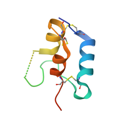
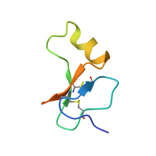
    Insulin-like growth factors (IGFs) are key regulators of cell proliferation, differentiation and transformation, and are thus pivotal in cancer, especially breast, prostate and colon neoplasms. They are also important in many neurological and bone disorders. Their potent mitogenic and anti-apoptotic actions depend primarily on their availability to bind to the cell surface IGF-I receptor. In circulation and interstitial fluids, IGFs are largely unavailable as they are tightly associated with IGF-binding proteins (IGFBPs) and are released after IGFBP proteolysis. Here we report the 2.1 A crystal structure of the complex of IGF-I bound to the N-terminal IGF-binding domain of IGFBP-5 (mini-IGFBP-5), a prototype interaction for all N-terminal domains of the IGFBP family. The principal interactions in the complex comprise interlaced hydrophobic side chains that protrude from both IGF-I and the IGFBP-5 fragment and a surrounding network of polar interactions. A solvent-exposed hydrophobic patch is located on the IGF-I pole opposite to the mini-IGFBP-5 binding region and marks the IGF-I receptor binding site.


  • Organizational Affiliation
    • Max Planck Institute for Biochemistry, D-82152 Martinsried, Germany.

Macromolecule Content 

  • Total Structure Weight: 13.72 kDa 
  • Atom Count: 809 
  • Modeled Residue Count: 99 
  • Deposited Residue Count: 124 
  • Unique protein chains: 2

Macromolecules

Find similar proteins by:|  3D Structure
Entity ID: 1
MoleculeChains  Sequence LengthOrganismDetailsImage
INSULIN-LIKE GROWTH FACTOR IA70Homo sapiensMutation(s): 0 
UniProt & NIH Common Fund Data Resources
Find proteins for P05019 (Homo sapiens)
Explore P05019 
Go to UniProtKB:  P05019
PHAROS:  P05019
GTEx:  ENSG00000017427 
Entity Groups
Sequence Clusters30% Identity50% Identity70% Identity90% Identity95% Identity100% Identity
UniProt GroupP05019
Sequence Annotations
Expand
Reference Sequence
Find similar proteins by:|  3D Structure
Entity ID: 2
MoleculeChains  Sequence LengthOrganismDetailsImage
INSULIN-LIKE GROWTH FACTOR BINDING PROTEIN 554Homo sapiensMutation(s): 0 
UniProt & NIH Common Fund Data Resources
Find proteins for P24593 (Homo sapiens)
Explore P24593 
Go to UniProtKB:  P24593
PHAROS:  P24593
GTEx:  ENSG00000115461 
Entity Groups
Sequence Clusters30% Identity50% Identity70% Identity90% Identity95% Identity100% Identity
UniProt GroupP24593
Sequence Annotations
Expand
Reference Sequence

Experimental Data & Validation

Experimental Data

  • Method: X-RAY DIFFRACTION
  • Resolution: 2.10 Å
  • R-Value Free:  0.262 (Depositor) 
  • R-Value Work:  0.218 (Depositor) 
  • R-Value Observed: 0.218 (Depositor) 
Space Group: P 21 3
Unit Cell:
Length ( Å )Angle ( ˚ )
a = 74.385α = 90
b = 74.385β = 90
c = 74.385γ = 90
Software Package:
Software NamePurpose
CNSrefinement
MOSFLMdata reduction
SCALAdata scaling

Structure Validation

View Full Validation Report


Entry History 

Deposition Data

Revision History  (Full details and data files)

  • Version 1.0: 2002-05-16
    Type: Initial release
  • Version 1.1: 2011-05-08
    Changes: Version format compliance
  • Version 1.2: 2011-07-13
    Changes: Version format compliance
  • Version 1.3: 2024-11-06
    Changes: Data collection, Database references, Structure summary