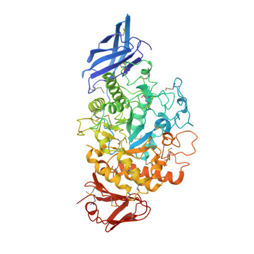
Covalent and Three-Dimensional Structure of the Cyclodextrinase from Flavobacterium Sp. No. 92.
Fritzsche, H.B., Schwede, T., Schulz, G.E.(2003) Eur J Biochem 270: 2332-2341
- PubMed: 12752453
- DOI: https://doi.org/10.1046/j.1432-1033.2003.03603.x
- Primary Citation of Related Structures:
1H3G - PubMed Abstract:
Starting with oligopeptide sequences and using PCR, the gene of the cyclodextrinase from Flavobacterium sp. no. 92 was derived from the genomic DNA. The gene was sequenced and expressed in Escherichia coli; the gene product was purified and crystallized. An X-ray diffraction analysis using seleno-methionines with multiwavelength anomalous diffraction techniques yielded the refined 3D structure at 2.1 A resolution. The enzyme hydrolyzes alpha(1,4)-glycosidic bonds of cyclodextrins and linear malto-oligosaccharides. It belongs to the glycosylhydrolase family no. 13 and has a chain fold similar to that of alpha-amylases, cyclodextrin glycosyltransferases, and other cyclodextrinases. In contrast with most family members but in agreement with other cyclodextrinases, the enzyme contains an additional characteristic N-terminal domain of about 100 residues. This domain participates in the formation of a putative D2-symmetric tetramer but not in cyclodextrin binding at the active center as observed with the other cyclodextrinases. Moreover, the domain is located at a position quite different from that of the other cyclodextrinases. Whether oligomerization facilitates the cyclodextrin deformation required for hydrolysis is discussed.
- Institut für Organische Chemie und Biochemie, Albert-Ludwigs-Universität, Freiburg im Breisgau, Germany.
Organizational Affiliation:


