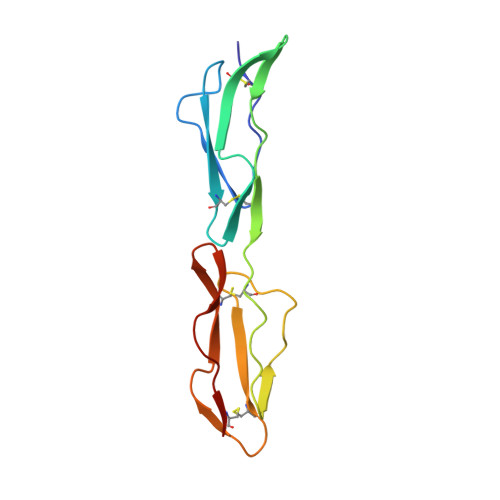
1H04

Human CD55 domains 3 & 4


Experimental Data Snapshot

  • Method: X-RAY DIFFRACTION
  • Resolution: 2.00 Å
  • R-Value Free: 0.259 
  • R-Value Work: 0.214 
  • R-Value Observed: 0.214 

wwPDB Validation   3D Report Full Report


This is version 1.2 of the entry. See complete history


Literature

Mapping Cd55 Function. The Structure of Two Pathogen-Binding Domains at 1.7 A

Williams, P.Chaudhry, Y.Goodfellow, I.Billington, J.Powell, R.Spiller, O.Evans, D.J.Lea, S.M.

(2003) J Biol Chem 278: 10691

  • DOI: https://doi.org/10.1074/jbc.M212561200
  • Primary Citation of Related Structures:  
    1H03, 1H04, 1H2P, 1H2Q, 1UOT

  • PubMed Abstract: 
  • Decay-accelerating factor (CD55), a regulator of the alternative and classical pathways of complement activation, is expressed on all serum-exposed cells. It is used by pathogens, including many enteroviruses and uropathogenic Escherichia coli, as a receptor prior to infection ...

    Decay-accelerating factor (CD55), a regulator of the alternative and classical pathways of complement activation, is expressed on all serum-exposed cells. It is used by pathogens, including many enteroviruses and uropathogenic Escherichia coli, as a receptor prior to infection. We describe the x-ray structure of a pathogen-binding fragment of human CD55 at 1.7 A resolution containing two of the three domains required for regulation of human complement. We have used mutagenesis to map biological functions onto the molecule; decay-accelerating activity maps to a single face of the molecule, whereas bacterial and viral pathogens recognize a variety of different sites on CD55.


    Related Citations: 
    • Crystallization and Preliminary X-Ray Diffraction Analysis of a Biologically Active Fragment of Cd55
      Lea, S.M., Powell, R., Evans, D.J.
      (1999) Acta Crystallogr D Biol Crystallogr D55: 1198
    • Determination of the Affinity and Kinetic Constants for the Interaction between the Human Virus Echovirus 11 and its Cellular Receptor, Cd55
      Lea, S.M., Powell, R., Mckee, T., Evans, D.J., Brown, D.J., Stuart, D.I., Van Der Merwe, A.
      (1998) J Biol Chem 273: 30443

    Organizational Affiliation

    Laboratory of Molecular Biophysics, Department of Biochemistry, University of Oxford, United Kingdom.


Macromolecules
Find similar proteins by:  (by identity cutoff)  |  3D Structure
Entity ID: 1
MoleculeChainsSequence LengthOrganismDetailsImage
COMPLEMENT DECAY-ACCELERATING FACTORA [auth P]125Homo sapiensMutation(s): 0 
Gene Names: CD55CRDAF
UniProt & NIH Common Fund Data Resources
Find proteins for P08174 (Homo sapiens)
Explore P08174 
Go to UniProtKB:  P08174
PHAROS:  P08174
Entity Groups  
Sequence Clusters30% Identity50% Identity70% Identity90% Identity95% Identity100% Identity
UniProt GroupP08174
Protein Feature View
Expand
  • Reference Sequence
Small Molecules
Ligands 1 Unique
IDChainsName / Formula / InChI Key2D Diagram3D Interactions
NI
Query on NI

Download Ideal Coordinates CCD File 
B [auth P]NICKEL (II) ION
Ni
VEQPNABPJHWNSG-UHFFFAOYSA-N
 Ligand Interaction
Experimental Data & Validation

Experimental Data

  • Method: X-RAY DIFFRACTION
  • Resolution: 2.00 Å
  • R-Value Free: 0.259 
  • R-Value Work: 0.214 
  • R-Value Observed: 0.214 
  • Space Group: C 1 2 1
Unit Cell:
Length ( Å )Angle ( ˚ )
a = 125.7α = 90
b = 20.4β = 111.5
c = 52.7γ = 90
Software Package:
Software NamePurpose
CNSrefinement
DENZOdata reduction
SCALEPACKdata scaling
EPMRphasing

Structure Validation

View Full Validation Report


Entry History 

Deposition Data

Revision History  (Full details and data files)

  • Version 1.0: 2003-03-20
    Type: Initial release
  • Version 1.1: 2011-05-08
    Changes: Version format compliance
  • Version 1.2: 2011-07-13
    Changes: Version format compliance