1GZJ

Structure of Thermoascus aurantiacus family 5 endoglucanase


Experimental Data Snapshot

  • Method: X-RAY DIFFRACTION
  • Resolution: 1.62 Å
  • R-Value Free: 0.177 
  • R-Value Work: 0.159 
  • R-Value Observed: 0.159 

wwPDB Validation   3D Report Full Report


This is version 1.4 of the entry. See complete history


Literature

The 1.62 A Structure of Thermoascus Aurantiacus Endoglucanase: Completing the Structural Picture of Subfamilies in Glycoside Hydrolase Family 5

Lo Leggio, L.Larsen, S.

(2002) FEBS Lett 523: 103-108

  • DOI: https://doi.org/10.1016/s0014-5793(02)02954-x
  • Primary Citation of Related Structures:  
    1GZJ

  • PubMed Abstract: 

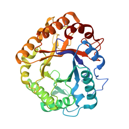
    The crystal structure of Thermoascus aurantiacus endoglucanase (Cel5A), a family 5 glycoside hydrolase, has been determined to 1.62 A resolution by multiple isomorphous replacement with anomalous scattering. It is the first report of a structure in the subfamily to which Cel5A belongs. Cel5A consists solely of a catalytic module with compact eight-fold beta/alpha barrel architecture. The length of the tryptophan-rich substrate binding groove suggests the presence of substrate binding subsites -4 to +3. Structural comparison shows that two glycines are completely conserved in the family, in addition to the two catalytic glutamates and six other conserved residues previously identified. Gly 44 in particular is part of a type IV C-terminal helix capping motif, whose disruption is likely to affect the position of an essential conserved arginine. One aromatic residue (Trp 170 in Cel5A), not conserved in term of sequence, is nonetheless spatially conserved in the substrate binding groove. Its role might be to force the bend that occurs in the polysaccharide chain on binding, thus favoring substrate distortion at subsite -1.


  • Organizational Affiliation

    Centre for Crystallographic Studies, Department of Chemistry, Chemical Institute, University of Copenhagen, Universitetsparken 5, DK-2100, Copenhagen, Denmark. leila@ccs.ki.ku.dk


Macromolecules
Find similar proteins by:  (by identity cutoff)  |  3D Structure
Entity ID: 1
MoleculeChains Sequence LengthOrganismDetailsImage
EGI
A, B
304Thermoascus aurantiacusMutation(s): 0 
EC: 3.2.1.4
UniProt
Find proteins for Q8TG26 (Thermoascus aurantiacus)
Explore Q8TG26 
Go to UniProtKB:  Q8TG26
Entity Groups  
Sequence Clusters30% Identity50% Identity70% Identity90% Identity95% Identity100% Identity
UniProt GroupQ8TG26
Sequence Annotations
Expand
  • Reference Sequence
Experimental Data & Validation

Experimental Data

  • Method: X-RAY DIFFRACTION
  • Resolution: 1.62 Å
  • R-Value Free: 0.177 
  • R-Value Work: 0.159 
  • R-Value Observed: 0.159 
  • Space Group: P 21 21 21
Unit Cell:
Length ( Å )Angle ( ˚ )
a = 76.51α = 90
b = 85.85β = 90
c = 89.64γ = 90
Software Package:
Software NamePurpose
CNSrefinement
DENZOdata reduction
Agrovatadata scaling
ROTAVATAdata scaling
SHARPphasing
ARPphasing
DMphasing

Structure Validation

View Full Validation Report


Entry History 

Deposition Data

Revision History  (Full details and data files)

  • Version 1.0: 2002-08-06
    Type: Initial release
  • Version 1.1: 2011-05-08
    Changes: Version format compliance
  • Version 1.2: 2011-07-13
    Changes: Version format compliance
  • Version 1.3: 2018-06-13
    Changes: Data collection, Database references, Source and taxonomy, Structure summary
  • Version 1.4: 2019-05-08
    Changes: Data collection, Experimental preparation