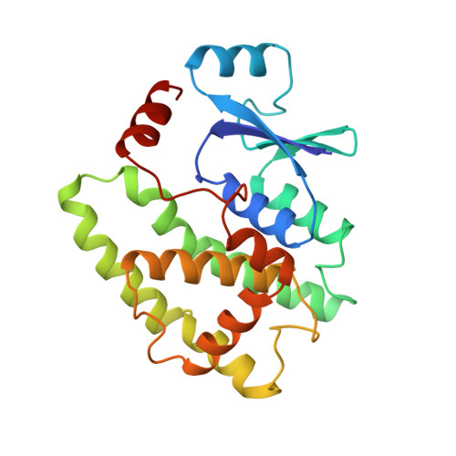
Human glutathione transferase A4-4 crystal structures and mutagenesis reveal the basis of high catalytic efficiency with toxic lipid peroxidation products
Bruns, C.M., Hubatsch, I., Ridderstrom, M., Mannervik, B., Tainer, J.A.(1999) J Mol Biol 288: 427-439
- PubMed: 10329152 Search on PubMed
- DOI: https://doi.org/10.1006/jmbi.1999.2697
- Primary Citation Related Structures:
1GUL, 1GUM - PubMed Abstract:
The oxidation of lipids and cell membranes generates cytotoxic compounds implicated in the etiology of aging, cancer, atherosclerosis, neurodegenerative diseases, and other illnesses. Glutathione transferase (GST) A4-4 is a key component in the defense against the products of this oxidative stress because, unlike other Alpha class GSTs, GST A4-4 shows high catalytic activity with lipid peroxidation products such as 4-hydroxynon-2-enal (HNE). The crystal structure of human apo GST A4-4 unexpectedly possesses an ordered C-terminal alpha-helix, despite the absence of any ligand. The structure of human GST A4-4 in complex with the inhibitor S-(2-iodobenzyl) glutathione reveals key features of the electrophilic substrate-binding pocket which confer specificity toward HNE. Three structural modules form the binding site for electrophilic substrates and thereby govern substrate selectivity: the beta1-alpha1 loop, the end of the alpha4 helix, and the C-terminal alpha9 helix. A few residue changes in GST A4-4 result in alpha9 taking over a predominant role in ligand specificity from the N-terminal loop region important for GST A1-1. Thus, the C-terminal helix alpha9 in GST A4-4 provides pre-existing ligand complementarity rather than acting as a flexible cap as observed in other GST structures. Hydrophobic residues in the alpha9 helix, differing from those in the closely related GST A1-1, delineate a hydrophobic specificity canyon for the binding of lipid peroxidation products. The role of residue Tyr212 as a key catalytic residue, suggested by the crystal structure of the inhibitor complex, is confirmed by mutagenesis results. Tyr212 is positioned to interact with the aldehyde group of the substrate and polarize it for reaction. Tyr212 also coopts part of the binding cleft ordinarily formed by the N-terminal substrate recognition region in the homologous enzyme GST A1-1 to reveal an evolutionary swapping of function between different recognition elements. A structural model of catalysis is presented based on these results.
- Department of Molecular Biology MB4, Skaggs Institute for Chemical Biology, The Scripps Research Institute, 10550 N. Torrey Pines Road, La Jolla, CA, 92037, USA.
Organizational Affiliation: