1GS0

Crystal structure of the catalytic fragment of murine poly(ADP-ribose) polymerase-2


Experimental Data Snapshot

  • Method: X-RAY DIFFRACTION
  • Resolution: 2.80 Å
  • R-Value Free: 0.268 
  • R-Value Work: 0.244 
  • R-Value Observed: 0.244 

wwPDB Validation   3D Report Full Report


This is version 1.3 of the entry. See complete history


Literature

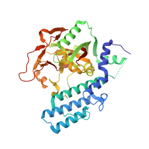
Crystal Structure of the Catalytic Fragment of Murine Poly(Adp-Ribose) Polymerase-2

Oliver, A.W.Ame, J.C.Roe, S.M.Good, V.De Murcia, G.Pearl, L.H.

(2004) Nucleic Acids Res 32: 456

  • DOI: https://doi.org/10.1093/nar/gkh215
  • Primary Citation of Related Structures:  
    1GS0

  • PubMed Abstract: 

    Poly(ADP-ribose) polymerase-1 (PARP-1) has become an important pharmacological target in the treatment of cancer due to its cellular role as a 'DNA-strand break sensor', which leads in part to resistance to some existing chemo- and radiological treatments. Inhibitors have now been developed which prevent PARP-1 from synthesizing poly(ADP-ribose) in response to DNA-breaks and potentiate the cytotoxicity of DNA damaging agents. However, with the recent discoveries of PARP-2, which has a similar DNA-damage dependent catalytic activity, and additional members containing the 'PARP catalytic' signature, the isoform selectivity and resultant pharmacological effects of existing inhibitors are brought into question. We present here the crystal structure of the catalytic fragment of murine PARP-2, at 2.8 A resolution, and compare this to the catalytic fragment of PARP-1, with an emphasis on providing a possible framework for rational drug design in order to develop future isoform-specific inhibitors.


  • Organizational Affiliation

    Cancer Research UK DNA Repair Enzyme Group, Section of Structural Biology, The Institute of Cancer Research, Chester Beatty Laboratories, 237 Fulham Road, London SW3 6JB, UK.


Macromolecules
Find similar proteins by:  (by identity cutoff)  |  3D Structure
Entity ID: 1
MoleculeChains Sequence LengthOrganismDetailsImage
POLY (ADP-RIBOSE) POLYMERASE-2
A, B
351Mus musculusMutation(s): 0 
EC: 2.4.2.30
UniProt
Find proteins for O88554 (Mus musculus)
Explore O88554 
Go to UniProtKB:  O88554
Entity Groups  
Sequence Clusters30% Identity50% Identity70% Identity90% Identity95% Identity100% Identity
UniProt GroupO88554
Sequence Annotations
Expand
  • Reference Sequence
Experimental Data & Validation

Experimental Data

  • Method: X-RAY DIFFRACTION
  • Resolution: 2.80 Å
  • R-Value Free: 0.268 
  • R-Value Work: 0.244 
  • R-Value Observed: 0.244 
  • Space Group: P 21 21 21
Unit Cell:
Length ( Å )Angle ( ˚ )
a = 83.65α = 90
b = 85.82β = 90
c = 139.46γ = 90
Software Package:
Software NamePurpose
CNSrefinement
MOSFLMdata reduction
SCALAdata scaling
AMoREphasing

Structure Validation

View Full Validation Report


Entry History 

Deposition Data

Revision History  (Full details and data files)

  • Version 1.0: 2002-12-19
    Type: Initial release
  • Version 1.1: 2011-05-08
    Changes: Version format compliance
  • Version 1.2: 2011-07-13
    Changes: Version format compliance
  • Version 1.3: 2023-12-13
    Changes: Data collection, Database references, Other, Refinement description