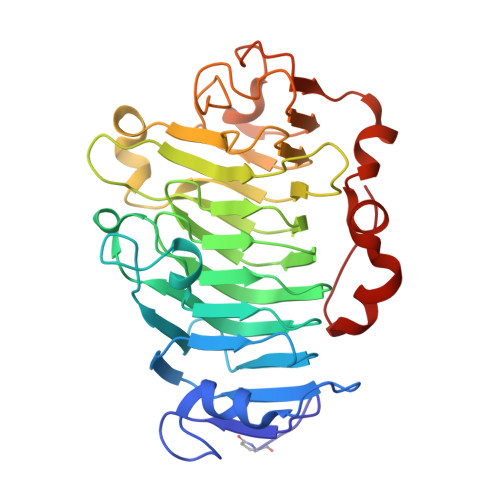
1GQ8

Pectin methylesterase from Carrot


Experimental Data Snapshot

  • Method: X-RAY DIFFRACTION
  • Resolution: 1.75 Å
  • R-Value Free: 0.193 
  • R-Value Work: 0.181 
  • R-Value Observed: 0.181 

Starting Model: experimental
View more details

wwPDB Validation   3D Report Full Report


This is version 2.1 of the entry. See complete history


Literature

Crystal Structure of Plant Pectin Methylesterase

Johansson, K.El-Ahmad, M.Friemann, R.Jornvall, H.Markovic, O.Eklund, H.

(2002) FEBS Lett 514: 243

  • DOI: https://doi.org/10.1016/s0014-5793(02)02372-4
  • Primary Citation of Related Structures:  
    1GQ8

  • PubMed Abstract: 

    Pectin is a principal component in the primary cell wall of plants. During cell development, pectin is modified by pectin methylesterases to give different properties to the cell wall. This report describes the first crystal structure of a plant pectin methylesterase. The beta-helical structure embodies a central cleft, lined by several aromatic residues, that has been deduced to be suitable for pectin binding. The active site is found at the center of this cleft where Asp157 is suggested to act as the nucleophile, Asp136 as an acid/base and Gln113/Gln135 to form an anion hole to stabilize the transition state.


  • Organizational Affiliation

    Department of Molecular Biology, Swedish University of Agricultural Sciences, S-751 24 Uppsala, Sweden.


Macromolecules
Find similar proteins by:  (by identity cutoff)  |  3D Structure
Entity ID: 1
MoleculeChains Sequence LengthOrganismDetailsImage
PECTINESTERASE319Daucus carotaMutation(s): 0 
EC: 3.1.1.11
UniProt
Find proteins for P83218 (Daucus carota)
Explore P83218 
Go to UniProtKB:  P83218
Entity Groups  
Sequence Clusters30% Identity50% Identity70% Identity90% Identity95% Identity100% Identity
UniProt GroupP83218
Sequence Annotations
Expand
  • Reference Sequence
Small Molecules
Modified Residues  1 Unique
IDChains TypeFormula2D DiagramParent
PCA
Query on PCA
A
L-PEPTIDE LINKINGC5 H7 N O3GLN
Experimental Data & Validation

Experimental Data

  • Method: X-RAY DIFFRACTION
  • Resolution: 1.75 Å
  • R-Value Free: 0.193 
  • R-Value Work: 0.181 
  • R-Value Observed: 0.181 
  • Space Group: P 21 21 2
Unit Cell:
Length ( Å )Angle ( ˚ )
a = 77.69α = 90
b = 89.41β = 90
c = 49.5γ = 90
Software Package:
Software NamePurpose
CNSrefinement
MOSFLMdata reduction
SCALAdata scaling
AMoREphasing

Structure Validation

View Full Validation Report


Entry History 

Deposition Data

Revision History  (Full details and data files)

  • Version 1.0: 2002-04-18
    Type: Initial release
  • Version 1.1: 2011-09-14
    Changes: Database references, Derived calculations, Non-polymer description, Other, Refinement description, Structure summary, Version format compliance
  • Version 1.2: 2018-01-17
    Changes: Data collection
  • Version 2.0: 2020-03-11
    Changes: Derived calculations, Other, Polymer sequence
  • Version 2.1: 2023-12-13
    Changes: Data collection, Database references, Refinement description