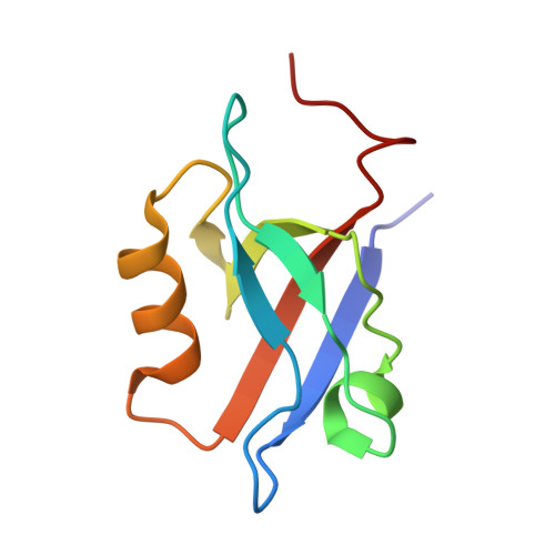
Structural Determinants of the Na+/H+ Exchanger Regulatory Factor Interaction with the Beta 2 Adrenergic and Platelet-Derived Growth Factor Receptors
Karthikeyan, S., Leung, T., Ladias, J.A.A.(2002) J Biological Chem 277: 18973
- PubMed: 11882663
- DOI: https://doi.org/10.1074/jbc.M201507200
- Primary Citation of Related Structures:
1GQ4, 1GQ5 - PubMed Abstract:
The Na(+)/H(+) exchanger regulatory factor (NHERF) binds through its PDZ1 domain to the carboxyl-terminal sequences NDSLL and EDSFL of the beta(2) adrenergic receptor (beta(2)AR) and platelet-derived growth factor receptor, respectively, and plays a critical role in the membrane localization and physiological regulation of these receptors. The crystal structures of the human NHERF PDZ1 domain bound to the sequences NDSLL and EDSFL have been determined at 1.9- and 2.2-A resolution, respectively. The beta(2)AR and platelet-derived growth factor receptor ligands insert into the PDZ1 binding pocket by a beta-sheet augmentation process and are stabilized by largely similar networks of hydrogen bonds and hydrophobic contacts. In the PDZ1-beta(2)AR complex, the side chain of asparagine at position -4 in the beta(2)AR peptide forms two additional hydrogen bonds with Gly(30) of PDZ1, which contribute to the higher affinity of this interaction. Remarkably, both complexes are further stabilized by hydrophobic interactions involving the side chains of the penultimate amino acids of the peptide ligands, whereas the PDZ1 residues Asn(22) and Glu(43) undergo conformational changes to accommodate these side chains. These results provide structural insights into the mechanisms by which different side chains at the position -1 of peptide ligands interact with PDZ domains and contribute to the affinity of the PDZ-ligand interaction.
- Molecular Medicine Laboratory and Macromolecular Crystallography Unit, Division of Experimental Medicine, Harvard Institutes of Medicine, Harvard Medical School, Boston, Massachusetts 02115, USA.
Organizational Affiliation:


