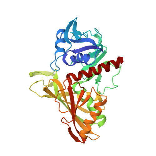
Studies of asymmetry in the three-dimensional structure of lobster D-glyceraldehyde-3-phosphate dehydrogenase.
Moras, D., Olsen, K.W., Sabesan, M.N., Buehner, M., Ford, G.C., Rossmann, M.G.(1975) J Biological Chem 250: 9137-9162
- PubMed: 127793
- DOI: https://doi.org/10.2210/pdb1gpd/pdb
- Primary Citation of Related Structures:
1GPD - PubMed Abstract:
An improved electron density map of lobster holo-D-glyceraldehyde-3-phosphate dehydrogenase has been computed to 2.9 A resolution based on two heavy atom isomorphous derivatives. This has been averaged only over the Q molecular 2-fold axis, which is known to be exact in the human holoenzyme. The map showed possible asymmetry between the subunits in which the active centers are closely related across the R axis (that is, between the red and green or between the yellow and blue subunits). A difference map between the electron density of citrate and sulfate-soaked crystals gave further evidence for possible asymmetry. The major differences of electron density between R axis-related subunits appear around the active center and suggest the following interpretations. 1. The conformation of the adenine about the glycosidic bond is the more frequently observed anti with a C-2' endo conformation for the ribose ring in the red and yellow subunits, but is probably syn with a C-3' endo conformation in the green and blue subunits.2. The adenine ribose has its 3'-hydroxyl group hydrogen-bonded to a main chain carbonyl group in the red and yellow subunits but not in the green and blue subunits, as a consequence of the differing ribose conformations. 3. Cysteine-149 is more closely associated with histidine-176 in the green and blue subunits, and appears nearer the nicotinamide in the red and yellow subunits.


