1GOL

COORDINATES OF RAT MAP KINASE ERK2 WITH AN ARGININE MUTATION AT POSITION 52


Experimental Data Snapshot

  • Method: X-RAY DIFFRACTION
  • Resolution: 2.80 Å

wwPDB Validation   3D Report Full Report


This is version 1.3 of the entry. See complete history


Literature

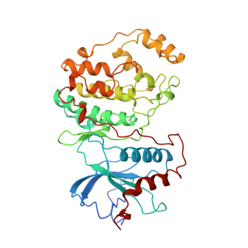
Mutation of position 52 in ERK2 creates a nonproductive binding mode for adenosine 5'-triphosphate.

Robinson, M.J.Harkins, P.C.Zhang, J.Baer, R.Haycock, J.W.Cobb, M.H.Goldsmith, E.J.

(1996) Biochemistry 35: 5641-5646

  • DOI: https://doi.org/10.1021/bi952723e
  • Primary Citation of Related Structures:  
    1GOL

  • PubMed Abstract: 
  • Among the protein kinases, an absolutely conserved lysine in subdomain II is required for high catalytic activity. This lysine is known to interact with the substrate ATP, but otherwise its role is not well understood. We have used biochemical and structural methods to investigate the function of this lysine (K52) in phosphoryl transfer reactions catalyzed by the MAP kinase ERK2 ...

    Among the protein kinases, an absolutely conserved lysine in subdomain II is required for high catalytic activity. This lysine is known to interact with the substrate ATP, but otherwise its role is not well understood. We have used biochemical and structural methods to investigate the function of this lysine (K52) in phosphoryl transfer reactions catalyzed by the MAP kinase ERK2. The kinetic properties of activated wild-type ERK2 and K52 mutants were examined using the oncoprotein TAL2, myelin basic protein, and a designed synthetic peptide as substrates. The catalytic activities of K52R and K52A ERK2 were lower than that of wild-type ERK2, primarily as a consequence of reductions in kcat. Further, there was little difference in Km for ATP, but the Km,app for peptide substrate was higher for the K52 mutants. The three-dimensional structure of unphosphorylated K52R ERK2 in the absence and presence of bound ATP was determined and compared with the structure of unphosphorylated wild-type ERK2. ATP adopted a well-defined but distinct binding mode in K52R ERK2 compared to the binding mode in the wild-type enzyme. The structural and kinetic data show that mutation of K52 created a nonproductive binding mode for ATP and suggest that K52 is essential for orienting ATP for catalysis.


    Related Citations: 
    • Activity of the Map Kinase Erk2 is Controlled by a Flexible Surface Loop
      Zhang, J., Zhang, F., Ebert, D., Cobb, M.H., Goldsmith, E.J.
      (1995) Structure 3: 299
    • Atomic Structure of the Map Kinase Erk2 at 2.3 A Resolution
      Zhang, F., Strand, A., Robbins, D., Cobb, M.H., Goldsmith, E.J.
      (1994) Nature 367: 704
    • Crystallization and Preliminary X-Ray Studies of Extracellular Signal-Regulated Kinase-2/Map Kinase with an Incorporated His-Tag
      Zhang, F., Robbins, D.J., Cobb, M.H., Goldsmith, E.J.
      (1993) J Mol Biol 233: 550

    Organizational Affiliation

    Department of Pharmacology, University of Texas Southwestern Medical Center, Dallas 75235, USA.


Macromolecules
Find similar proteins by:  (by identity cutoff)  |  3D Structure
Entity ID: 1
MoleculeChainsSequence LengthOrganismDetailsImage
EXTRACELLULAR REGULATED KINASE 2364Rattus norvegicusMutation(s): 1 
Gene Names: Erk2Mapk1MapkPrkm1
EC: 2.7.1 (PDB Primary Data), 2.7.11.24 (UniProt)
UniProt
Find proteins for P63086 (Rattus norvegicus)
Explore P63086 
Go to UniProtKB:  P63086
Entity Groups  
Sequence Clusters30% Identity50% Identity70% Identity90% Identity95% Identity100% Identity
UniProt GroupP63086
Protein Feature View
Expand
  • Reference Sequence
Small Molecules
Ligands 2 Unique
IDChainsName / Formula / InChI Key2D Diagram3D Interactions
ATP
Query on ATP

Download Ideal Coordinates CCD File 
C [auth A]ADENOSINE-5'-TRIPHOSPHATE
C10 H16 N5 O13 P3
ZKHQWZAMYRWXGA-KQYNXXCUSA-N
 Ligand Interaction
MG
Query on MG

Download Ideal Coordinates CCD File 
B [auth A]MAGNESIUM ION
Mg
JLVVSXFLKOJNIY-UHFFFAOYSA-N
 Ligand Interaction
Experimental Data & Validation

Experimental Data

  • Method: X-RAY DIFFRACTION
  • Resolution: 2.80 Å
  • Space Group: P 1 21 1
Unit Cell:
Length ( Å )Angle ( ˚ )
a = 49.32α = 90
b = 71.42β = 109.75
c = 61.25γ = 90
Software Package:
Software NamePurpose
DENZOdata reduction
SCALEPACKdata scaling

Structure Validation

View Full Validation Report


Entry History 

Deposition Data

Revision History  (Full details and data files)

  • Version 1.0: 1997-03-12
    Type: Initial release
  • Version 1.1: 2008-03-24
    Changes: Version format compliance
  • Version 1.2: 2011-07-13
    Changes: Version format compliance
  • Version 1.3: 2021-11-03
    Changes: Database references, Derived calculations, Other