1GER

THE STRUCTURE OF GLUTATHIONE REDUCTASE FROM ESCHERICHIA COLI AT 1.86 ANGSTROMS RESOLUTION: COMPARISON WITH THE ENZYME FROM HUMAN ERYTHROCYTES


Experimental Data Snapshot

  • Method: X-RAY DIFFRACTION
  • Resolution: 1.86 Å
  • R-Value Work: 0.168 
  • R-Value Observed: 0.168 

wwPDB Validation   3D Report Full Report


This is version 1.2 of the entry. See complete history


Literature

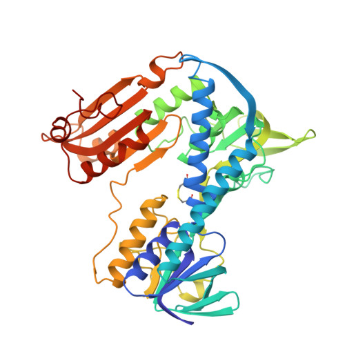
Structure of glutathione reductase from Escherichia coli at 1.86 A resolution: comparison with the enzyme from human erythrocytes.

Mittl, P.R.Schulz, G.E.

(1994) Protein Sci 3: 799-809

  • DOI: https://doi.org/10.1002/pro.5560030509
  • Primary Citation of Related Structures:  
    1GER

  • PubMed Abstract: 

    The crystal structure of the dimeric flavoenzyme glutathione reductase from Escherichia coli was determined and refined to an R-factor of 16.8% at 1.86 A resolution. The molecular 2-fold axis of the dimer is local but very close to a possible crystallographic 2-fold axis; the slight asymmetry could be rationalized from the packing contacts. The 2 crystallographically independent subunits of the dimer are virtually identical, yielding no structural clue on possible cooperativity. The structure was compared with the well-known structure of the homologous enzyme from human erythrocytes with 52% sequence identity. Significant differences were found at the dimer interface, where the human enzyme has a disulfide bridge, whereas the E. coli enzyme has an antiparallel beta-sheet connecting the subunits. The differences at the glutathione binding site and in particular a deformation caused by a Leu-Ile exchange indicate why the E. coli enzyme accepts trypanothione much better than the human enzyme. The reported structure provides a frame for explaining numerous published engineering results in detail and for guiding further ones.


  • Organizational Affiliation

    Institut für Organische Chemie und Biochemie, Albert-Ludwigs-Universität, Freiburg, Germany.


Macromolecules
Find similar proteins by:  (by identity cutoff)  |  3D Structure
Entity ID: 1
MoleculeChains Sequence LengthOrganismDetailsImage
GLUTATHIONE REDUCTASE
A, B
450Escherichia coliMutation(s): 0 
EC: 1.6.4.2 (PDB Primary Data), 1.8.1.7 (UniProt)
UniProt
Find proteins for P06715 (Escherichia coli (strain K12))
Explore P06715 
Go to UniProtKB:  P06715
Entity Groups  
Sequence Clusters30% Identity50% Identity70% Identity90% Identity95% Identity100% Identity
UniProt GroupP06715
Sequence Annotations
Expand
  • Reference Sequence
Small Molecules
Ligands 1 Unique
IDChains Name / Formula / InChI Key2D Diagram3D Interactions
FAD
Query on FAD

Download Ideal Coordinates CCD File 
C [auth A],
D [auth B]
FLAVIN-ADENINE DINUCLEOTIDE
C27 H33 N9 O15 P2
VWWQXMAJTJZDQX-UYBVJOGSSA-N
Experimental Data & Validation

Experimental Data

  • Method: X-RAY DIFFRACTION
  • Resolution: 1.86 Å
  • R-Value Work: 0.168 
  • R-Value Observed: 0.168 
  • Space Group: P 1 1 21
Unit Cell:
Length ( Å )Angle ( ˚ )
a = 120.5α = 90
b = 73.6β = 90
c = 60.5γ = 83
Software Package:
Software NamePurpose
X-PLORmodel building
X-PLORrefinement
X-PLORphasing

Structure Validation

View Full Validation Report


Entry History 

Deposition Data

Revision History  (Full details and data files)

  • Version 1.0: 1994-11-01
    Type: Initial release
  • Version 1.1: 2008-03-24
    Changes: Version format compliance
  • Version 1.2: 2011-07-13
    Changes: Version format compliance