1GBT

STRUCTURE OF AN ACYL-ENZYME INTERMEDIATE DURING CATALYSIS: (GUANIDINOBENZOYL) TRYPSIN


Experimental Data Snapshot

  • Method: X-RAY DIFFRACTION
  • Resolution: 2.00 Å
  • R-Value Observed: 0.162 

wwPDB Validation   3D Report Full Report


This is version 1.2 of the entry. See complete history


Literature

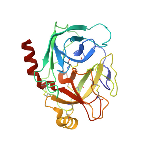
Structure of an acyl-enzyme intermediate during catalysis: (guanidinobenzoyl)trypsin.

Mangel, W.F.Singer, P.T.Cyr, D.M.Umland, T.C.Toledo, D.L.Stroud, R.M.Pflugrath, J.W.Sweet, R.M.

(1990) Biochemistry 29: 8351-8357

  • DOI: https://doi.org/10.1021/bi00488a022
  • Primary Citation of Related Structures:  
    1GBT

  • PubMed Abstract: 
  • The crystal and molecular structure of trypsin at a transiently stable intermediate step during catalysis has been determined by X-ray diffraction methods. Bovine trypsin cleaved the substrate p-nitrophenyl p-guanidinobenzoate during crystallization under conditions in which the acyl-enzyme intermediate, (guanidinobenzoyl)trypsin, was stable ...

    The crystal and molecular structure of trypsin at a transiently stable intermediate step during catalysis has been determined by X-ray diffraction methods. Bovine trypsin cleaved the substrate p-nitrophenyl p-guanidinobenzoate during crystallization under conditions in which the acyl-enzyme intermediate, (guanidinobenzoyl)trypsin, was stable. Orthorhombic crystals formed in space group P2(1)2(1)2(1), with a = 63.74, b = 63.54, and c = 68.93 A. This is a crystal form of bovine trypsin for which a molecular structure has not been reported. Diffraction data were measured with a FAST (Enraf Nonius) diffractometer. The structure was refined to a crystallographic residual of R = 0.16 for data in the resolution range 7.0-2.0 A. The refined model of (guanidinobenzoyl)trypsin provides insight into the structural basis for its slow rate of deacylation, which in solution at 25 degrees C and pH 7.4 exhibits a t1/2 of 12 h. In addition to the rotation of the Ser-195 hydroxyl away from His-157, C beta of Ser-195 moves 0.7 A toward Asp-189 at the bottom of the active site, with respect to the native structure. This allows formation of energetically favorable H bonds and an ion pair between the carboxylate of Asp-189 and the guanidino group of the substrate. This movement is dictated by the rigidity of the aromatic ring in guanidinobenzoate--model-building indicates that this should not occur when arginine, with its more flexible aliphatic backbone, forms the ester bond with Ser-195. As a consequence, highly ordered water molecules in the active site are no longer close enough to the scissile ester bond to serve as potential nucleophiles for hydrolysis.(ABSTRACT TRUNCATED AT 250 WORDS)


    Organizational Affiliation

    Biology Department, Brookhaven National Laboratory, Upton, New York 11973.


Macromolecules
Find similar proteins by:  (by identity cutoff)  |  3D Structure
Entity ID: 1
MoleculeChainsSequence LengthOrganismDetailsImage
BETA-TRYPSIN223Bos taurusMutation(s): 0 
Gene Names: PRSS1TRP1TRY1TRYP1
EC: 3.4.21.4
UniProt
Find proteins for P00760 (Bos taurus)
Explore P00760 
Go to UniProtKB:  P00760
Entity Groups  
Sequence Clusters30% Identity50% Identity70% Identity90% Identity95% Identity100% Identity
UniProt GroupP00760
Protein Feature View
Expand
  • Reference Sequence
Small Molecules
Ligands 3 Unique
IDChainsName / Formula / InChI Key2D Diagram3D Interactions
GBS
Query on GBS

Download Ideal Coordinates CCD File 
E [auth A]4-carbamimidamidobenzoic acid
C8 H9 N3 O2
SXTSBZBQQRIYCU-UHFFFAOYSA-N
 Ligand Interaction
SO4
Query on SO4

Download Ideal Coordinates CCD File 
C [auth A],
D [auth A]
SULFATE ION
O4 S
QAOWNCQODCNURD-UHFFFAOYSA-L
 Ligand Interaction
CA
Query on CA

Download Ideal Coordinates CCD File 
B [auth A]CALCIUM ION
Ca
BHPQYMZQTOCNFJ-UHFFFAOYSA-N
 Ligand Interaction
Binding Affinity Annotations 
IDSourceBinding Affinity
GBS PDBBind:  1GBT Ki: 1.00e+7 (nM) from 1 assay(s)
Experimental Data & Validation

Experimental Data

  • Method: X-RAY DIFFRACTION
  • Resolution: 2.00 Å
  • R-Value Observed: 0.162 
  • Space Group: P 21 21 21
Unit Cell:
Length ( Å )Angle ( ˚ )
a = 63.74α = 90
b = 63.54β = 90
c = 68.93γ = 90
Software Package:
Software NamePurpose
TNTrefinement

Structure Validation

View Full Validation Report


Entry History 

Deposition Data

Revision History  (Full details and data files)

  • Version 1.0: 1994-01-31
    Type: Initial release
  • Version 1.1: 2008-03-24
    Changes: Version format compliance
  • Version 1.2: 2011-07-13
    Changes: Version format compliance