1G73 | pdb_00001g73

CRYSTAL STRUCTURE OF SMAC BOUND TO XIAP-BIR3 DOMAIN


Experimental Data Snapshot

  • Method: X-RAY DIFFRACTION
  • Resolution: 2.00 Å
  • R-Value Free: 
    0.268 (Depositor) 
  • R-Value Work: 
    0.227 (Depositor) 
  • R-Value Observed: 
    0.227 (Depositor) 

wwPDB Validation 3D Report Full Report

Validation slider image for 1G73

This is version 1.4 of the entry. See complete history

Literature

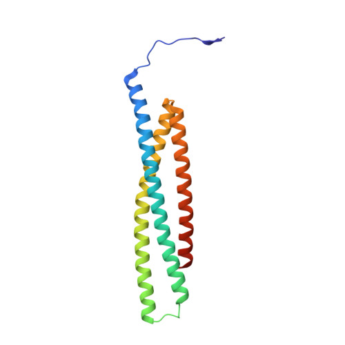
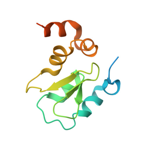
Structural basis of IAP recognition by Smac/DIABLO.

Wu, G.Chai, J.Suber, T.L.Wu, J.W.Du, C.Wang, X.Shi, Y.

(2000) Nature 408: 1008-1012

  • DOI: https://doi.org/10.1038/35050012
  • Primary Citation Related Structures: 
    1G73

  • PubMed Abstract: 

    Apoptosis is an essential process in the development and homeostasis of all metazoans. The inhibitor-of-apoptosis (IAP) proteins suppress cell death by inhibiting the activity of caspases; this inhibition is performed by the zinc-binding BIR domains of the IAP proteins. The mitochondrial protein Smac/DIABLO promotes apoptosis by eliminating the inhibitory effect of IAPs through physical interactions. Amino-terminal sequences in Smac/DIABLO are required for this function, as mutation of the very first amino acid leads to loss of interaction with IAPs and concomitant loss of Smac/DIABLO function. Here we report the high-resolution crystal structure of Smac/DIABLO complexed with the third BIR domain (BIR3) of XIAP. Our results show that the N-terminal four residues (Ala-Val-Pro-Ile) in Smac/DIABLO recognize a surface groove on BIR3, with the first residue Ala binding a hydrophobic pocket and making five hydrogen bonds to neighbouring residues on BIR3. These observations provide a structural explanation for the roles of the Smac N terminus as well as the conserved N-terminal sequences in the Drosophila proteins Hid/Grim/Reaper. In conjunction with other observations, our results reveal how Smac may relieve IAP inhibition of caspase-9 activity. In addition to explaining a number of biological observations, our structural analysis identifies potential targets for drug screening.


  • Organizational Affiliation
    • Department of Molecular Biology, Princeton University, New Jersey 08544, USA.

Macromolecule Content 

  • Total Structure Weight: 64.21 kDa 
  • Atom Count: 4,010 
  • Modeled Residue Count: 505 
  • Deposited Residue Count: 566 
  • Unique protein chains: 2

Macromolecules

Find similar proteins by:|  3D Structure
Entity ID: 1
MoleculeChains  Sequence LengthOrganismDetailsImage
SECOND MITOCHONDRIA-DERIVED ACTIVATOR OF CASPASES
A, B
162Homo sapiensMutation(s): 1 
Gene Names: SMAC
UniProt & NIH Common Fund Data Resources
Find proteins for Q9NR28 (Homo sapiens)
Explore Q9NR28 
Go to UniProtKB:  Q9NR28
PHAROS:  Q9NR28
GTEx:  ENSG00000184047 
Entity Groups
Sequence Clusters30% Identity50% Identity70% Identity90% Identity95% Identity100% Identity
UniProt GroupQ9NR28
Sequence Annotations
Expand
Reference Sequence
Find similar proteins by:|  3D Structure
Entity ID: 2
MoleculeChains  Sequence LengthOrganismDetailsImage
INHIBITORS OF APOPTOSIS-LIKE PROTEIN ILP
C, D
121Homo sapiensMutation(s): 0 
EC: 2.3.2.27
UniProt & NIH Common Fund Data Resources
Find proteins for P98170 (Homo sapiens)
Explore P98170 
Go to UniProtKB:  P98170
PHAROS:  P98170
GTEx:  ENSG00000101966 
Entity Groups
Sequence Clusters30% Identity50% Identity70% Identity90% Identity95% Identity100% Identity
UniProt GroupP98170
Sequence Annotations
Expand
Reference Sequence

Small Molecules

Experimental Data & Validation

Experimental Data

  • Method: X-RAY DIFFRACTION
  • Resolution: 2.00 Å
  • R-Value Free:  0.268 (Depositor) 
  • R-Value Work:  0.227 (Depositor) 
  • R-Value Observed: 0.227 (Depositor) 
Space Group: P 1
Unit Cell:
Length ( Å )Angle ( ˚ )
a = 48.1α = 100
b = 52.9β = 104.1
c = 67.1γ = 94
Software Package:
Software NamePurpose
MLPHAREphasing
X-PLORrefinement
DENZOdata reduction
SCALEPACKdata scaling

Structure Validation

View Full Validation Report


Entry History 

Deposition Data

Revision History  (Full details and data files)

  • Version 1.0: 2001-01-10
    Type: Initial release
  • Version 1.1: 2008-04-27
    Changes: Version format compliance
  • Version 1.2: 2011-07-13
    Changes: Version format compliance
  • Version 1.3: 2021-10-27
    Changes: Database references, Derived calculations
  • Version 1.4: 2024-02-07
    Changes: Data collection