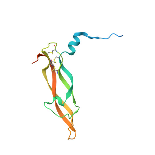
1FZV

THE CRYSTAL STRUCTURE OF HUMAN PLACENTA GROWTH FACTOR-1 (PLGF-1), AN ANGIOGENIC PROTEIN AT 2.0A RESOLUTION


Experimental Data Snapshot

  • Method: X-RAY DIFFRACTION
  • Resolution: 2.00 Å
  • R-Value Free: 0.247 
  • R-Value Work: 0.216 
  • R-Value Observed: 0.216 

wwPDB Validation   3D Report Full Report


This is version 1.4 of the entry. See complete history


Literature

The crystal structure of human placenta growth factor-1 (PlGF-1), an angiogenic protein, at 2.0 A resolution.

Iyer, S.Leonidas, D.D.Swaminathan, G.J.Maglione, D.Battisti, M.Tucci, M.Persico, M.G.Acharya, K.R.

(2001) J Biol Chem 276: 12153-12161

  • DOI: https://doi.org/10.1074/jbc.M008055200
  • Primary Citation of Related Structures:  
    1FZV

  • PubMed Abstract: 

    The angiogenic molecule placenta growth factor (PlGF) is a member of the cysteine-knot family of growth factors. In this study, a mature isoform of the human PlGF protein, PlGF-1, was crystallized as a homodimer in the crystallographic asymmetric unit, and its crystal structure was elucidated at 2.0 A resolution. The overall structure of PlGF-1 is similar to that of vascular endothelial growth factor (VEGF) with which it shares 42% amino acid sequence identity. Based on structural and biochemical data, we have mapped several important residues on the PlGF-1 molecule that are involved in recognition of the fms-like tyrosine kinase receptor (Flt-1, also known as VEGFR-1). We propose a model for the association of PlGF-1 and Flt-1 domain 2 with precise shape complementarity, consider the relevance of this assembly for PlGF-1 signal transduction, and provide a structural basis for altered specificity of this molecule.


  • Organizational Affiliation

    Department of Biology and Biochemistry, University of Bath, Claverton Down, Bath BA2 7AY, United Kingdom.


Macromolecules
Find similar proteins by:  (by identity cutoff)  |  3D Structure
Entity ID: 1
MoleculeChains Sequence LengthOrganismDetailsImage
PLACENTA GROWTH FACTOR
A, B
132Homo sapiensMutation(s): 0 
UniProt & NIH Common Fund Data Resources
Find proteins for P49763 (Homo sapiens)
Explore P49763 
Go to UniProtKB:  P49763
PHAROS:  P49763
GTEx:  ENSG00000119630 
Entity Groups  
Sequence Clusters30% Identity50% Identity70% Identity90% Identity95% Identity100% Identity
UniProt GroupP49763
Sequence Annotations
Expand
  • Reference Sequence
Small Molecules
Ligands 1 Unique
IDChains Name / Formula / InChI Key2D Diagram3D Interactions
MPD
Query on MPD

Download Ideal Coordinates CCD File 
C [auth A],
D [auth B]
(4S)-2-METHYL-2,4-PENTANEDIOL
C6 H14 O2
SVTBMSDMJJWYQN-YFKPBYRVSA-N
Experimental Data & Validation

Experimental Data

  • Method: X-RAY DIFFRACTION
  • Resolution: 2.00 Å
  • R-Value Free: 0.247 
  • R-Value Work: 0.216 
  • R-Value Observed: 0.216 
  • Space Group: P 43
Unit Cell:
Length ( Å )Angle ( ˚ )
a = 62.6α = 90
b = 62.6β = 90
c = 84.107γ = 90
Software Package:
Software NamePurpose
DENZOdata reduction
SCALEPACKdata scaling
AMoREphasing
CNSrefinement

Structure Validation

View Full Validation Report


Entry History 

Deposition Data

Revision History  (Full details and data files)

  • Version 1.0: 2001-05-09
    Type: Initial release
  • Version 1.1: 2008-04-27
    Changes: Version format compliance
  • Version 1.2: 2011-07-13
    Changes: Version format compliance
  • Version 1.3: 2018-01-31
    Changes: Experimental preparation
  • Version 1.4: 2022-12-21
    Changes: Database references, Derived calculations