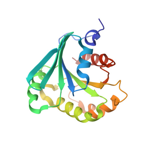
1FZQ

CRYSTAL STRUCTURE OF MURINE ARL3-GDP


Experimental Data Snapshot

  • Method: X-RAY DIFFRACTION
  • Resolution: 1.70 Å
  • R-Value Free: 0.235 
  • R-Value Work: 0.213 
  • R-Value Observed: 0.215 

Starting Model: experimental
View more details

wwPDB Validation   3D Report Full Report


Ligand Structure Quality Assessment 


This is version 1.3 of the entry. See complete history


Literature

Structural and biochemical properties show ARL3-GDP as a distinct GTP binding protein.

Hillig, R.C.Hanzal-Bayer, M.Linari, M.Becker, J.Wittinghofer, A.Renault, L.

(2000) Structure 8: 1239-1245

  • DOI: https://doi.org/10.1016/s0969-2126(00)00531-1
  • Primary Citation of Related Structures:  
    1FZQ

  • PubMed Abstract: 

    Based on sequence similarities, Arf-like (ARL) proteins have been assigned to the Arf subfamily of the superfamily of Ras-related GTP binding proteins. They have been identified in several isoforms in a wide variety of species. Their cellular function is unclear, but they are proposed to regulate intracellular transport. The 1.7 A crystal structure of murine ARL3-GDP provides a first insight into the structural features of this subgroup of Ar proteins. The N-terminal extension of ARL3 folds into an elongated loop region that is hydrophobically anchored onto the surface by burying 1440 A2. The features observed suggest that ARL3 releases its N terminus and undergoes a beta sheet register shift upon the binding of GTP. The structure and kinetic experiments with fluorescent mGDP demonstrate that tight GDP (but not GTP) binding is achieved in the absence of a magnesium ion. This is due to a lysine residue in the active site, close to the canonical Mg2+ site found in other GTP binding proteins. This is a distinct feature separating ARL2 and ARL3 from Arf proteins. The disturbed magnesium binding site and the independence of GDP coordination from the presence of Mg2+ separate ARL2 and ARL3 from Arf proteins. The D sheet register shift, which is similar to that of Arf, that is observed in the present structure, along with the postulated release of the N-terminal extension and the concomitant exposure of a patch of conserved hydrophobic residues in this region suggest that ARL proteins might be localized to target membranes upon exchange of GDP to GTP. Contrary to the situation in Arf, however, the conformational change to ARL-GTP does not require the presence of membranes and might thus be energetically unfavored. Together with the very low affinity described for the interaction of ARL3 with Mg-GTP, this suggests that ARL protein activation requires the presence of effectors stabilizing the GTP coordination rather than guanine nucleotide exchange factors (GEFs).


  • Organizational Affiliation

    Max-Planck-Institut für Molekulare Physiologie, Abteilung Strukturelle Biologie, Dortmund, Germany.


Macromolecules
Find similar proteins by:  (by identity cutoff)  |  3D Structure
Entity ID: 1
MoleculeChains Sequence LengthOrganismDetailsImage
ADP-RIBOSYLATION FACTOR-LIKE PROTEIN 3181Mus musculusMutation(s): 0 
Membrane Entity: Yes 
UniProt
Find proteins for Q9WUL7 (Mus musculus)
Explore Q9WUL7 
Go to UniProtKB:  Q9WUL7
Entity Groups  
Sequence Clusters30% Identity50% Identity70% Identity90% Identity95% Identity100% Identity
UniProt GroupQ9WUL7
Sequence Annotations
Expand
  • Reference Sequence
Small Molecules
Ligands 4 Unique
IDChains Name / Formula / InChI Key2D Diagram3D Interactions
GDP
Query on GDP

Download Ideal Coordinates CCD File 
F [auth A]GUANOSINE-5'-DIPHOSPHATE
C10 H15 N5 O11 P2
QGWNDRXFNXRZMB-UUOKFMHZSA-N
MES
Query on MES

Download Ideal Coordinates CCD File 
E [auth A]2-(N-MORPHOLINO)-ETHANESULFONIC ACID
C6 H13 N O4 S
SXGZJKUKBWWHRA-UHFFFAOYSA-N
SO4
Query on SO4

Download Ideal Coordinates CCD File 
B [auth A],
C [auth A]
SULFATE ION
O4 S
QAOWNCQODCNURD-UHFFFAOYSA-L
NH4
Query on NH4

Download Ideal Coordinates CCD File 
D [auth A]AMMONIUM ION
H4 N
QGZKDVFQNNGYKY-UHFFFAOYSA-O
Binding Affinity Annotations 
IDSourceBinding Affinity
GDP PDBBind:  1FZQ Kd: 24 (nM) from 1 assay(s)
Experimental Data & Validation

Experimental Data

  • Method: X-RAY DIFFRACTION
  • Resolution: 1.70 Å
  • R-Value Free: 0.235 
  • R-Value Work: 0.213 
  • R-Value Observed: 0.215 
  • Space Group: P 21 21 2
Unit Cell:
Length ( Å )Angle ( ˚ )
a = 74.421α = 90
b = 64.583β = 90
c = 40.386γ = 90
Software Package:
Software NamePurpose
AMoREphasing
CNSrefinement
DENZOdata reduction
SCALEPACKdata scaling

Structure Validation

View Full Validation Report


Ligand Structure Quality Assessment 


Entry History 

Deposition Data

Revision History  (Full details and data files)

  • Version 1.0: 2000-12-06
    Type: Initial release
  • Version 1.1: 2008-04-27
    Changes: Version format compliance
  • Version 1.2: 2011-07-13
    Changes: Version format compliance
  • Version 1.3: 2023-08-09
    Changes: Data collection, Database references, Derived calculations, Refinement description