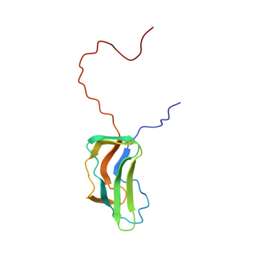
1FYC

INNER LIPOYL DOMAIN FROM HUMAN PYRUVATE DEHYDROGENASE (PDH) COMPLEX, NMR, 1 STRUCTURE


Experimental Data Snapshot

  • Method: SOLUTION NMR
  • Conformers Calculated: 80 
  • Conformers Submitted: 
  • Selection Criteria: MEAN STRUCTURE FROM 26 CHOSEN HAVING LEAST RESTRAINT VIOLATION 

wwPDB Validation   3D Report Full Report


This is version 1.4 of the entry. See complete history


Literature

Three-dimensional structure of the major autoantigen in primary biliary cirrhosis.

Howard, M.J.Fuller, C.Broadhurst, R.W.Perham, R.N.Tang, J.G.Quinn, J.Diamond, A.G.Yeaman, S.J.

(1998) Gastroenterology 115: 139-146

  • DOI: https://doi.org/10.1016/s0016-5085(98)70375-0
  • Primary Citation of Related Structures:  
    1FYC

  • PubMed Abstract: 

    Primary biliary cirrhosis (PBC) is a chronic cholestatic liver disease characterized by the presence of antimitochondrial autoantibodies in patients' serum. The major autoantigen, recognized by antibodies from > 95% of patients with PBC, has been identified as the E2 component (E2p) of the pyruvate dehydrogenase multienzyme complex. Immunodominant sites on E2p have been localized to the inner of the two lipoyl domains, where the essential cofactor lipoic acid is attached covalently. The aim of this study was to determine the three-dimensional structure of the inner lipoyl domain of human E2p. The domain was expressed in Escherichia coli; after purification, its structure was analyzed using nuclear magnetic resonance spectroscopy. The structure of the lipoyl domain from human E2p was determined, and the implications of the structure for autoimmune recognition were assessed. Knowledge of the structure further defines the major epitope and may help in the design of antigen-specific immunotherapy for treatment of PBC.


  • Organizational Affiliation

    Department of Biochemistry, Cambridge Centre for Molecular Recognition, University of Cambridge, England.


Macromolecules
Find similar proteins by:  (by identity cutoff)  |  3D Structure
Entity ID: 1
MoleculeChains Sequence LengthOrganismDetailsImage
DIHYDROLIPOAMIDE ACETYLTRANSFERASE (E2P)106Homo sapiensMutation(s): 0 
EC: 2.3.1.12
UniProt & NIH Common Fund Data Resources
Find proteins for P10515 (Homo sapiens)
Explore P10515 
Go to UniProtKB:  P10515
PHAROS:  P10515
GTEx:  ENSG00000150768 
Entity Groups  
Sequence Clusters30% Identity50% Identity70% Identity90% Identity95% Identity100% Identity
UniProt GroupP10515
Sequence Annotations
Expand
  • Reference Sequence
Experimental Data & Validation

Experimental Data

  • Method: SOLUTION NMR
  • Conformers Calculated: 80 
  • Conformers Submitted: 
  • Selection Criteria: MEAN STRUCTURE FROM 26 CHOSEN HAVING LEAST RESTRAINT VIOLATION 

Structure Validation

View Full Validation Report


Entry History 

Deposition Data

Revision History  (Full details and data files)

  • Version 1.0: 1997-09-04
    Type: Initial release
  • Version 1.1: 2008-03-24
    Changes: Version format compliance
  • Version 1.2: 2011-07-13
    Changes: Version format compliance
  • Version 1.3: 2017-11-29
    Changes: Derived calculations, Other
  • Version 1.4: 2024-05-01
    Changes: Data collection, Database references