1FSE | pdb_00001fse

CRYSTAL STRUCTURE OF THE BACILLUS SUBTILIS REGULATORY PROTEIN GERE


Experimental Data Snapshot

  • Method: X-RAY DIFFRACTION
  • Resolution: 2.05 Å
  • R-Value Free: 
    0.272 (Depositor) 
  • R-Value Work: 
    0.214 (Depositor) 

wwPDB Validation 3D Report Full Report

Validation slider image for 1FSE

This is version 1.5 of the entry. See complete history

Literature

Crystal structure of GerE, the ultimate transcriptional regulator of spore formation in Bacillus subtilis.

Ducros, V.M.Lewis, R.J.Verma, C.S.Dodson, E.J.Leonard, G.Turkenburg, J.P.Murshudov, G.N.Wilkinson, A.J.Brannigan, J.A.

(2001) J Mol Biology 306: 759-771

  • DOI: https://doi.org/10.1006/jmbi.2001.4443
  • Primary Citation Related Structures: 
    1FSE

  • PubMed Abstract: 

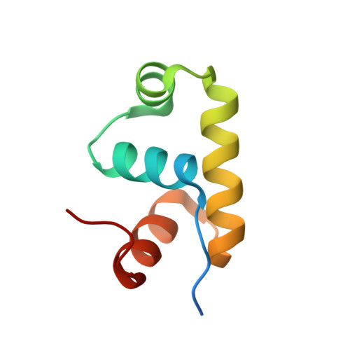
    The small, DNA-binding protein GerE regulates gene transcription in the terminally differentiated mother-cell compartment during late stages of sporulation in Bacillus subtilis. This versatile transcription factor shares sequence homology with the LuxR/FixJ/UhpA family of activators and modulates the expression of a number of genes, in particular those encoding the components of the coat that surrounds the mature spore. GerE orchestrates the final stages of coat deposition and maturation that lead to a spore with remarkable resistance properties but that must be responsive to low levels of germination signals. As this germination process is largely passive and can occur in the absence of de novo protein synthesis, the correct assembly of germination machinery, including germinant receptors and energy storage compounds, is crucial to the survival of the cell. The crystal structure of GerE has been solved at 2.05 A resolution using multi-wavelength anomalous dispersion techniques and reveals the nature of the GerE dimer. Each monomer comprises four alpha-helices, of which the central pair forms a helix-turn-helix DNA-binding motif. Implications for DNA-binding and the structural organisation of the LuxR/FixJ/UhpA family of transcription activator domains are discussed.


  • Organizational Affiliation
    • Structural Biology Laboratory Department of Chemistry, University of York, Heslington, YO10 5DD, UK.

Macromolecule Content 

  • Total Structure Weight: 52.28 kDa 
  • Atom Count: 3,484 
  • Modeled Residue Count: 384 
  • Deposited Residue Count: 444 
  • Unique protein chains: 1

Macromolecules

Find similar proteins by:|  3D Structure
Entity ID: 1
MoleculeChains  Sequence LengthOrganismDetailsImage
GERE
A, B, C, D, E
A, B, C, D, E, F
74Bacillus subtilisMutation(s): 0 
UniProt
Find proteins for P11470 (Bacillus subtilis (strain 168))
Explore P11470 
Go to UniProtKB:  P11470
Entity Groups
Sequence Clusters30% Identity50% Identity70% Identity90% Identity95% Identity100% Identity
UniProt GroupP11470
Sequence Annotations
Expand
Reference Sequence

Experimental Data & Validation

Experimental Data

  • Method: X-RAY DIFFRACTION
  • Resolution: 2.05 Å
  • R-Value Free:  0.272 (Depositor) 
  • R-Value Work:  0.214 (Depositor) 
Space Group: C 1 2 1
Unit Cell:
Length ( Å )Angle ( ˚ )
a = 109.019α = 90
b = 61.749β = 97.08
c = 71.743γ = 90
Software Package:
Software NamePurpose
MLPHAREphasing
REFMACrefinement
DENZOdata reduction
SCALEPACKdata scaling

Structure Validation

View Full Validation Report


Entry History 

Deposition Data

Revision History  (Full details and data files)

  • Version 1.0: 2001-03-21
    Type: Initial release
  • Version 1.1: 2008-04-27
    Changes: Version format compliance
  • Version 1.2: 2011-07-13
    Changes: Non-polymer description, Version format compliance
  • Version 1.3: 2018-01-31
    Changes: Database references
  • Version 1.4: 2018-02-14
    Changes: Experimental preparation
  • Version 1.5: 2024-02-07
    Changes: Data collection, Database references, Derived calculations