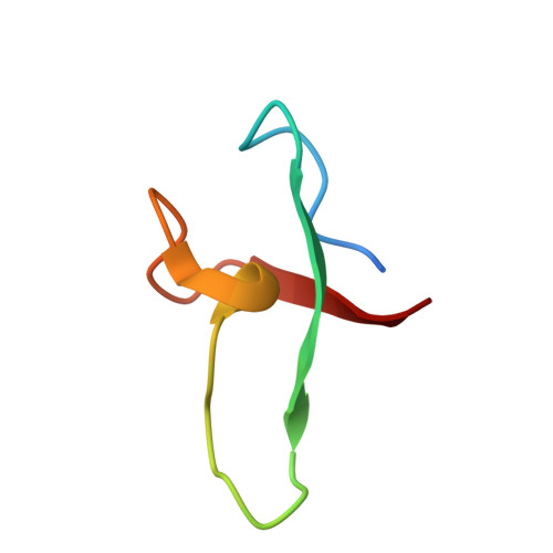
Novel topology of a zinc-binding domain from a protein involved in regulating early Xenopus development.
Borden, K.L., Lally, J.M., Martin, S.R., O'Reilly, N.J., Etkin, L.D., Freemont, P.S.(1995) EMBO J 14: 5947-5956
- PubMed: 8846787
- DOI: https://doi.org/10.1002/j.1460-2075.1995.tb00283.x
- Primary Citation of Related Structures:
1FRE - PubMed Abstract:
Xenopus nuclear factor XNF7, a maternally expressed protein, functions in patterning of the embryo. XNF7 contains a number of defined protein domains implicated in the regulation of some developmental processes. Among these is a tripartite motif comprising a zinc-binding RING finger and B-box domain next to a predicted alpha-helical coiled-coil domain. Interestingly, this motif is found in a variety of protein including several proto-oncoproteins. Here we describe the solution structure of the XNF7 B-box zinc-binding domain determined at physiological pH by 1H NMR methods. The B-box structure represents the first three-dimensional structure of this new motif and comprises a monomer have two beta-strands, two helical turns and three extended loop regions packed in a novel topology. The r.m.s. deviation for the best 18 structures is 1.15 A for backbone atoms and 1.94 A for all atoms. Structure calculations and biochemical data shows one zinc atom ligated in a Cys2-His2 tetrahedral arrangement. We have used mutant peptides to determine the metal ligation scheme which surprisingly shows that not all of the seven conserved cysteines/histidines in the B-box motif are involved in metal ligation. The B-box structure is not similar in tertiary fold to any other known zinc-binding motif.
- Laboratory of Molecular Structure, National Institute for Medical Research, London, UK.
Organizational Affiliation:


