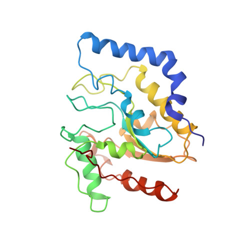
Stressing-out DNA? The contribution of serine-phosphodiester interactions in catalysis by uracil DNA glycosylase.
Werner, R.M., Jiang, Y.L., Gordley, R.G., Jagadeesh, G.J., Ladner, J.E., Xiao, G., Tordova, M., Gilliland, G.L., Stivers, J.T.(2000) Biochemistry 39: 12585-12594
- PubMed: 11027138
- DOI: https://doi.org/10.1021/bi001532v
- Primary Citation of Related Structures:
1FLZ - PubMed Abstract:
The DNA repair enzyme uracil DNA glycosylase (UDG) pinches the phosphodiester backbone of damaged DNA using the hydroxyl side chains of a conserved trio of serine residues, resulting in flipping of the deoxyuridine from the DNA helix into the enzyme active site. We have investigated the energetic role of these serine-phosphodiester interactions using the complementary approaches of crystallography, directed mutagenesis, and stereospecific phosphorothioate substitutions. A new crystal structure of UDG bound to 5'-HO-dUAAp-3' (which lacks the 5' phosphodiester group that interacts with the Ser88 pinching finger) shows that the glycosidic bond of dU has been cleaved, and that the enzyme has undergone the same specific clamping motion that brings key active site groups into position as previously observed in the structures of human UDG bound to large duplex DNA substrates. From this structure, it may be concluded that glycosidic bond cleavage and the induced fit conformational change in UDG can occur without the 5' pinching interaction. The S88A, S189A, and S192G "pinching" mutations exhibit 360-, 80-, and 21-fold damaging effects on k(cat)/K(m), respectively, while the S88A/S189A double mutant exhibits an 8200-fold damaging effect. A free energy analysis of the combined effects of nonbridging phosphorothioate substitution and mutation at these positions reveals the presence of a modest amount of strain energy between the compressed 5' and 3' phosphodiester groups flanking the bound uridine. Overall, these results indicate a role for these serine-phosphodiester interactions in uracil flipping and preorganization of the sugar ring into a reactive conformation. However, in contrast to a recent proposal [Parikh, S. S., et al. (2000) Proc Natl. Acad. Sci. 94, 5083], there is no evidence that conformational strain of the glycosidic bond induced by serine pinching plays a major role in the 10(12)-fold rate enhancement brought about by UDG.
- Center for Advanced Research in Biotechnology, University of Maryland Biotechnology Institute and National Institute for Standards and Technology, 9600 Gudelsky Drive, Rockville, Maryland 20850, USA.
Organizational Affiliation:


