1FJS | pdb_00001fjs

CRYSTAL STRUCTURE OF THE INHIBITOR ZK-807834 (CI-1031) COMPLEXED WITH FACTOR XA


Experimental Data Snapshot

  • Method: X-RAY DIFFRACTION
  • Resolution: 1.92 Å
  • R-Value Free: 
    0.256 (Depositor), 0.252 (DCC) 
  • R-Value Work: 
    0.196 (Depositor), 0.189 (DCC) 

Starting Model: experimental
View more details

wwPDB Validation 3D Report Full Report

Validation slider image for 1FJS

Ligand Structure Quality Assessment 


This is version 1.6 of the entry. See complete history

Literature

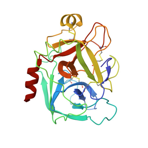
Preparation, characterization, and the crystal structure of the inhibitor ZK-807834 (CI-1031) complexed with factor Xa.

Adler, M.Davey, D.D.Phillips, G.B.Kim, S.H.Jancarik, J.Rumennik, G.Light, D.R.Whitlow, M.

(2000) Biochemistry 39: 12534-12542

  • DOI: https://doi.org/10.1021/bi001477q
  • Primary Citation Related Structures: 
    1FJS

  • PubMed Abstract: 

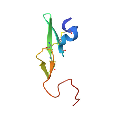
    Factor Xa plays a critical role in the formation of blood clots. This serine protease catalyzes the conversion of prothrombin to thrombin, the first joint step that links the intrinsic and extrinsic coagulation pathways. There is considerable interest in the development of factor Xa inhibitors for the intervention in thrombic diseases. This paper presents the structure of the inhibitor ZK-807834, also known as CI-1031, bound to factor Xa and provides the details of the protein purification and crystallization. Results from mass spectrometry indicate that the factor Xa underwent autolysis during crystallization and the first EGF-like domain was cleaved from the protein. The crystal structure of the complex shows that the amidine of ZK-807834 forms a salt bridge with Asp189 in the S1 pocket and the basic imidazoline fits snugly into the S4 site. The central pyridine ring provides a fairly rigid linker between these groups. This rigidity helps minimize entropic losses during binding. In addition, the structure reveals new interactions that were not found in the previous factor Xa/inhibitor complexes. ZK-807834 forms a strong hydrogen bond between an ionized 2-hydroxy group and Ser195 of factor Xa. There is also an aromatic ring-stacking interaction between the inhibitor and Trp215 in the S4 pocket. These interactions contribute to both the potency of this compound (K(I) = 0.11 nM) and the >2500-fold selectivity against homologous serine proteases such as trypsin.


  • Organizational Affiliation
    • Berlex Biosciences, 15049 San Pablo Avenue, P.O. Box 4099, Richmond, California 94804-0099, USA.

Macromolecule Content 

  • Total Structure Weight: 33.19 kDa 
  • Atom Count: 2,475 
  • Modeled Residue Count: 286 
  • Deposited Residue Count: 286 
  • Unique protein chains: 2

Macromolecules

Find similar proteins by:|  3D Structure
Entity ID: 1
MoleculeChains  Sequence LengthOrganismDetailsImage
COAGULATION FACTOR XA234Homo sapiensMutation(s): 0 
EC: 3.4.21.6
UniProt & NIH Common Fund Data Resources
Find proteins for P00742 (Homo sapiens)
Explore P00742 
Go to UniProtKB:  P00742
PHAROS:  P00742
GTEx:  ENSG00000126218 
Entity Groups
Sequence Clusters30% Identity50% Identity70% Identity90% Identity95% Identity100% Identity
UniProt GroupP00742
Sequence Annotations
Expand
Reference Sequence
Find similar proteins by:|  3D Structure
Entity ID: 2
MoleculeChains  Sequence LengthOrganismDetailsImage
COAGULATION FACTOR XAB [auth L]52Homo sapiensMutation(s): 0 
EC: 3.4.21.6
UniProt & NIH Common Fund Data Resources
Find proteins for P00742 (Homo sapiens)
Explore P00742 
Go to UniProtKB:  P00742
PHAROS:  P00742
GTEx:  ENSG00000126218 
Entity Groups
Sequence Clusters30% Identity50% Identity70% Identity90% Identity95% Identity100% Identity
UniProt GroupP00742
Sequence Annotations
Expand
Reference Sequence

Small Molecules

Ligands 4 Unique
IDChains Name / Formula / InChI Key2D Diagram3D Interactions
Z34

Query on Z34


Download:Ideal Coordinates CCD File
E [auth A]N-[2-[5-[AMINO(IMINO)METHYL]-2-HYDROXYPHENOXY]-3,5-DIFLUORO-6-[3-(4,5-DIHYDRO-1-METHYL-1H-IMIDAZOL-2-YL)PHENOXY]PYRIDIN-4-YL]-N-METHYLGLYCINE
C25 H24 F2 N6 O5
NPNSVNGQJGRSNR-UHFFFAOYSA-N
GOL

Query on GOL


Download:Ideal Coordinates CCD File
F [auth A]
G [auth A]
H [auth A]
I [auth A]
J [auth A]
F [auth A],
G [auth A],
H [auth A],
I [auth A],
J [auth A],
K [auth L]
GLYCEROL
C3 H8 O3
PEDCQBHIVMGVHV-UHFFFAOYSA-N
CA

Query on CA


Download:Ideal Coordinates CCD File
C [auth A]CALCIUM ION
Ca
BHPQYMZQTOCNFJ-UHFFFAOYSA-N
CL

Query on CL


Download:Ideal Coordinates CCD File
D [auth A]CHLORIDE ION
Cl
VEXZGXHMUGYJMC-UHFFFAOYSA-M
Binding Affinity Annotations 
IDSourceBinding Affinity
Z34 BindingDB:  1FJS Ki: 0.11 (nM) from 1 assay(s)

Experimental Data & Validation

Experimental Data

  • Method: X-RAY DIFFRACTION
  • Resolution: 1.92 Å
  • R-Value Free:  0.256 (Depositor), 0.252 (DCC) 
  • R-Value Work:  0.196 (Depositor), 0.189 (DCC) 
Space Group: P 21 21 21
Unit Cell:
Length ( Å )Angle ( ˚ )
a = 55.77α = 90
b = 71.96β = 90
c = 80.23γ = 90
Software Package:
Software NamePurpose
X-GENdata scaling
X-GENdata reduction
X-PLORmodel building
X-PLORrefinement
X-PLORphasing

Structure Validation

View Full Validation Report


Ligand Structure Quality Assessment 


Entry History 

Deposition Data

Revision History  (Full details and data files)

  • Version 1.0: 2000-10-04
    Type: Initial release
  • Version 1.1: 2008-04-27
    Changes: Version format compliance
  • Version 1.2: 2011-07-13
    Changes: Non-polymer description, Version format compliance
  • Version 1.3: 2017-10-04
    Changes: Advisory, Refinement description
  • Version 1.4: 2023-08-09
    Changes: Advisory, Data collection, Database references, Derived calculations, Refinement description
  • Version 1.5: 2024-03-13
    Changes: Source and taxonomy, Structure summary
  • Version 1.6: 2024-11-20
    Changes: Structure summary