1FBP | pdb_00001fbp

CRYSTAL STRUCTURE OF FRUCTOSE-1,6-BISPHOSPHATASE COMPLEXED WITH FRUCTOSE 6-PHOSPHATE, AMP, AND MAGNESIUM


Experimental Data Snapshot

  • Method: X-RAY DIFFRACTION
  • Resolution: 2.50 Å
  • R-Value Work: 
    0.215 (Depositor) 

wwPDB Validation 3D Report Full Report

Validation slider image for 1FBP

This is version 1.5 of the entry. See complete history

Literature

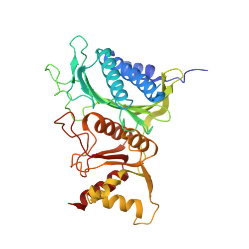
Crystal structure of fructose-1,6-bisphosphatase complexed with fructose 6-phosphate, AMP, and magnesium.

Ke, H.M.Zhang, Y.P.Lipscomb, W.N.

(1990) Proc Natl Acad Sci U S A 87: 5243-5247

  • DOI: https://doi.org/10.1073/pnas.87.14.5243
  • Primary Citation Related Structures: 
    1FBP

  • PubMed Abstract: 

    The crystal structure of fructose-1,6-bisphosphatase (EC 3.1.3.11) complexed with fructose 6-phosphate, AMP, and Mg2+ has been solved by the molecular replacement method and refined at 2.5-A resolution to a R factor of 0.215, with root-mean-square deviations of 0.013 A and 3.5 degrees for bond lengths and bond angles, respectively. No solvent molecules have been included in the refinement. This structure shows large quaternary and tertiary conformational changes from the structures of the unligated enzyme or its fructose 2,6-bisphosphate complex, but the secondary structures remain essentially the same. Dimer C3-C4 of the enzyme-fructose 6-phosphate-AMP-Mg2+ complex twists about 19 degrees relative to the same dimer of the enzyme-fructose 2,6-bisphosphate complex if their C1-C2 dimers are superimposed on one another. Nevertheless, many interfacial interactions between dimers of C1-C2 and C3-C4 are conserved after quaternary structure changes occur. Residues of the AMP domain (residues 6-200) show large migrations of C alpha atoms relative to barely significant positional changes of the FBP domain (residues 201-335).


  • Organizational Affiliation
    • Gibbs Chemical Laboratory, Harvard University, Cambridge, MA 02138.

Macromolecule Content 

  • Total Structure Weight: 74.27 kDa 
  • Atom Count: 4,940 
  • Modeled Residue Count: 634 
  • Deposited Residue Count: 670 
  • Unique protein chains: 1

Macromolecules

Find similar proteins by:|  3D Structure
Entity ID: 1
MoleculeChains  Sequence LengthOrganismDetailsImage
FRUCTOSE 1,6-BISPHOSPHATASE
A, B
335Sus scrofaMutation(s): 0 
EC: 3.1.3.11
UniProt
Find proteins for P00636 (Sus scrofa)
Explore P00636 
Go to UniProtKB:  P00636
Entity Groups
Sequence Clusters30% Identity50% Identity70% Identity90% Identity95% Identity100% Identity
UniProt GroupP00636
Sequence Annotations
Expand
Reference Sequence

Experimental Data & Validation

Experimental Data

  • Method: X-RAY DIFFRACTION
  • Resolution: 2.50 Å
  • R-Value Work:  0.215 (Depositor) 
Space Group: P 21 21 2
Unit Cell:
Length ( Å )Angle ( ˚ )
a = 61.6α = 90
b = 166.6β = 90
c = 80γ = 90
Software Package:
Software NamePurpose
X-PLORrefinement

Structure Validation

View Full Validation Report


Entry History 

Deposition Data

Revision History  (Full details and data files)

  • Version 1.0: 1992-04-15
    Type: Initial release
  • Version 1.1: 2008-03-24
    Changes: Version format compliance
  • Version 1.2: 2011-07-13
    Changes: Derived calculations, Version format compliance
  • Version 1.3: 2017-11-29
    Changes: Derived calculations, Other
  • Version 1.4: 2020-07-29
    Type: Remediation
    Reason: Carbohydrate remediation
    Changes: Data collection, Database references, Derived calculations, Structure summary
  • Version 1.5: 2024-12-25
    Changes: Advisory, Data collection, Database references, Derived calculations, Structure summary