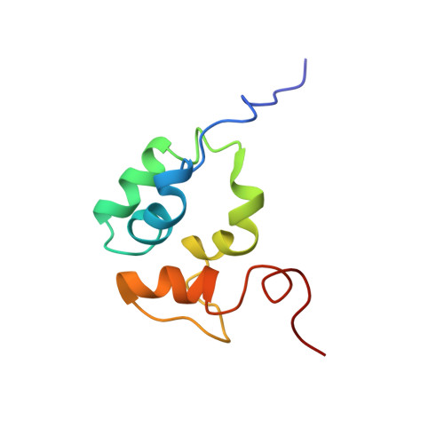
1F6V

SOLUTION STRUCTURE OF THE C TERMINAL OF MU B TRANSPOSITION PROTEIN


Experimental Data Snapshot

  • Method: SOLUTION NMR
  • Conformers Calculated: 100 
  • Conformers Submitted: 20 
  • Selection Criteria: structures with lowest energy and no restraint violations with phi psi angles in allowed regions 

wwPDB Validation   3D Report Full Report


This is version 1.4 of the entry. See complete history


Literature

The solution structure of the C-terminal domain of the Mu B transposition protein.

Hung, L.H.Chaconas, G.Shaw, G.S.

(2000) EMBO J 19: 5625-5634

  • DOI: https://doi.org/10.1093/emboj/19.21.5625
  • Primary Citation of Related Structures:  
    1F6V

  • PubMed Abstract: 

    Mu B is one of four proteins required for the strand transfer step of bacteriophage Mu DNA transposition and the only one where no high resolution structural data is available. Structural work on Mu B has been hampered primarily by solubility problems and its tendency to aggregate. We have overcome this problem by determination of the three-dimensional structure of the C-terminal domain of Mu B (B(223-312)) in 1.5 M NaCl using NMR spectroscopic methods. The structure of Mu B(223-312) comprises four helices (backbone r.m.s.d. 0.46 A) arranged in a loosely packed bundle and resembles that of the N-terminal region of the replication helicase, DnaB. This structural motif is likely to be involved in the inter-domainal regulation of ATPase activity for both Mu A and DnaB. The approach described here for structural determination in high salt may be generally applicable for proteins that do not crystallize and that are plagued by solubility problems at low ionic strength.


  • Organizational Affiliation

    Department of Biochemistry and McLaughlin Macromolecular Structure Facility, The University of Western Ontario, London, Ontario, Canada N6A 5C1. shaw@serena.biochem.uwo.ca


Macromolecules
Find similar proteins by:  (by identity cutoff)  |  3D Structure
Entity ID: 1
MoleculeChains Sequence LengthOrganismDetailsImage
DNA TRANSPOSITION PROTEIN91Muvirus muMutation(s): 0 
UniProt
Find proteins for P03763 (Escherichia phage Mu)
Explore P03763 
Go to UniProtKB:  P03763
Entity Groups  
Sequence Clusters30% Identity50% Identity70% Identity90% Identity95% Identity100% Identity
UniProt GroupP03763
Sequence Annotations
Expand
  • Reference Sequence
Experimental Data & Validation

Experimental Data

  • Method: SOLUTION NMR
  • Conformers Calculated: 100 
  • Conformers Submitted: 20 
  • Selection Criteria: structures with lowest energy and no restraint violations with phi psi angles in allowed regions 

Structure Validation

View Full Validation Report


Entry History 

Deposition Data

Revision History  (Full details and data files)

  • Version 1.0: 2000-11-08
    Type: Initial release
  • Version 1.1: 2008-04-27
    Changes: Version format compliance
  • Version 1.2: 2011-07-13
    Changes: Version format compliance
  • Version 1.3: 2017-02-01
    Changes: Structure summary
  • Version 1.4: 2024-05-01
    Changes: Data collection, Database references