1F6A

Structure of the human ige-fc bound to its high affinity receptor fc(epsilon)ri(alpha)


Experimental Data Snapshot

  • Method: X-RAY DIFFRACTION
  • Resolution: 3.50 Å
  • R-Value Free: 0.273 
  • R-Value Work: 0.254 
  • R-Value Observed: 0.254 

wwPDB Validation   3D Report Full Report


This is version 2.1 of the entry. See complete history


Literature

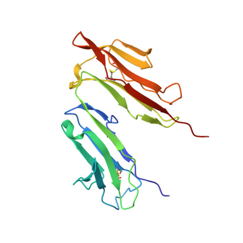
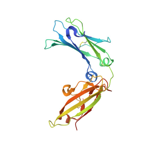
Structure of the Fc fragment of human IgE bound to its high-affinity receptor Fc (epsilon) RI (alpha).

Garman, S.C.Wurzburg, B.A.Tarchevskaya, S.S.Kinet, J.P.Jardetzky, T.S.

(2000) Nature 406: 259-266

  • DOI: https://doi.org/10.1038/35018500
  • Primary Citation of Related Structures:  
    1F6A

  • PubMed Abstract: 

    The initiation of immunoglobulin-E (IgE)-mediated allergic responses requires the binding of IgE antibody to its high-affinity receptor, Fc epsilonRI. Crosslinking of Fc epsilonRI initiates an intracellular signal transduction cascade that triggers the release of mediators of the allergic response. The interaction of the crystallizable fragment (Fc) of IgE (IgE-Fc) with Fc epsilonRI is a key recognition event of this process and involves the extracellular domains of the Fc epsilonRI alpha-chain. To understand the structural basis for this interaction, we have solved the crystal structure of the human IgE-Fc-Fc epsilonRI alpha complex to 3.5-A resolution. The crystal structure reveals that one receptor binds one dimeric IgE-Fc molecule asymmetrically through interactions at two sites, each involving one C epsilon3 domain of the IgE-Fc. The interaction of one receptor with the IgE-Fc blocks the binding of a second receptor, and features of this interaction are conserved in other members of the Fc receptor family. The structure suggests new approaches to inhibiting the binding of IgE to Fc epsilonRI for the treatment of allergy and asthma.


  • Organizational Affiliation

    Department of Biochemistry, Molecular Biology and Cell Biology, Northwestern University, Evanston, Illinois 60208, USA.


Macromolecules
Find similar proteins by:  (by identity cutoff)  |  3D Structure
Entity ID: 1
MoleculeChains Sequence LengthOrganismDetailsImage
HIGH AFFINITY IMMUNOGLOBULIN EPSILON RECEPTOR ALPHA-SUBUNIT176Homo sapiensMutation(s): 3 
UniProt & NIH Common Fund Data Resources
Find proteins for P12319 (Homo sapiens)
Explore P12319 
Go to UniProtKB:  P12319
PHAROS:  P12319
GTEx:  ENSG00000179639 
Entity Groups  
Sequence Clusters30% Identity50% Identity70% Identity90% Identity95% Identity100% Identity
UniProt GroupP12319
Sequence Annotations
Expand
  • Reference Sequence
Find similar proteins by:  (by identity cutoff)  |  3D Structure
Entity ID: 2
MoleculeChains Sequence LengthOrganismDetailsImage
IG EPSILON CHAIN C REGIONB,
C [auth D]
222Homo sapiensMutation(s): 0 
UniProt & NIH Common Fund Data Resources
Find proteins for P01854 (Homo sapiens)
Explore P01854 
Go to UniProtKB:  P01854
PHAROS:  P01854
GTEx:  ENSG00000211891 
Entity Groups  
Sequence Clusters30% Identity50% Identity70% Identity90% Identity95% Identity100% Identity
UniProt GroupP01854
Sequence Annotations
Expand
  • Reference Sequence
Oligosaccharides

Help

Entity ID: 3
MoleculeChains Length2D Diagram Glycosylation3D Interactions
alpha-D-mannopyranose-(1-4)-2-acetamido-2-deoxy-beta-D-glucopyranose-(1-4)-[alpha-L-fucopyranose-(1-6)]2-acetamido-2-deoxy-beta-D-glucopyranoseD [auth C]4N-Glycosylation
Glycosylation Resources
GlyTouCan:  G47477HI
GlyCosmos:  G47477HI
GlyGen:  G47477HI
Entity ID: 4
MoleculeChains Length2D Diagram Glycosylation3D Interactions
alpha-D-mannopyranose-(1-3)-[alpha-D-mannopyranose-(1-6)]alpha-D-mannopyranose-(1-4)-2-acetamido-2-deoxy-beta-D-glucopyranose-(1-4)-2-acetamido-2-deoxy-beta-D-glucopyranose
E
5N-Glycosylation
Glycosylation Resources
GlyTouCan:  G21381MC
GlyCosmos:  G21381MC
GlyGen:  G21381MC
Entity ID: 5
MoleculeChains Length2D Diagram Glycosylation3D Interactions
2-acetamido-2-deoxy-beta-D-glucopyranose-(1-4)-[alpha-L-fucopyranose-(1-6)]2-acetamido-2-deoxy-beta-D-glucopyranose
F
3N-Glycosylation
Glycosylation Resources
GlyTouCan:  G21290RB
GlyCosmos:  G21290RB
GlyGen:  G21290RB
Entity ID: 6
MoleculeChains Length2D Diagram Glycosylation3D Interactions
alpha-D-mannopyranose-(1-2)-alpha-D-mannopyranose-(1-6)-[alpha-D-mannopyranose-(1-3)]alpha-D-mannopyranose-(1-4)-2-acetamido-2-deoxy-beta-D-glucopyranose-(1-4)-2-acetamido-2-deoxy-beta-D-glucopyranose
G
6N-Glycosylation
Glycosylation Resources
GlyTouCan:  G08161XL
GlyCosmos:  G08161XL
GlyGen:  G08161XL
Entity ID: 7
MoleculeChains Length2D Diagram Glycosylation3D Interactions
alpha-D-mannopyranose-(1-4)-2-acetamido-2-deoxy-beta-D-glucopyranose-(1-4)-2-acetamido-2-deoxy-beta-D-glucopyranose
H
3N-Glycosylation
Glycosylation Resources
GlyTouCan:  G62182OO
GlyCosmos:  G62182OO
GlyGen:  G62182OO
Experimental Data & Validation

Experimental Data

  • Method: X-RAY DIFFRACTION
  • Resolution: 3.50 Å
  • R-Value Free: 0.273 
  • R-Value Work: 0.254 
  • R-Value Observed: 0.254 
  • Space Group: H 3 2
Unit Cell:
Length ( Å )Angle ( ˚ )
a = 192.8α = 90
b = 192.8β = 90
c = 302.4γ = 120
Software Package:
Software NamePurpose
DENZOdata reduction
SCALEPACKdata scaling
AMoREphasing
CNSrefinement

Structure Validation

View Full Validation Report


Entry History 

Deposition Data

Revision History  (Full details and data files)

  • Version 1.0: 2000-07-20
    Type: Initial release
  • Version 1.1: 2008-04-27
    Changes: Version format compliance
  • Version 1.2: 2011-07-13
    Changes: Non-polymer description, Version format compliance
  • Version 2.0: 2020-07-29
    Type: Remediation
    Reason: Carbohydrate remediation
    Changes: Advisory, Atomic model, Data collection, Database references, Derived calculations, Structure summary
  • Version 2.1: 2021-11-03
    Changes: Database references, Derived calculations, Structure summary