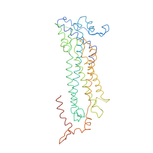
The crystal structure of adenylosuccinate lyase from Pyrobaculum aerophilum reveals an intracellular protein with three disulfide bonds.
Toth, E.A., Worby, C., Dixon, J.E., Goedken, E.R., Marqusee, S., Yeates, T.O.(2000) J Mol Biology 301: 433-450
- PubMed: 10926519 Search on PubMed
- DOI: https://doi.org/10.1006/jmbi.2000.3970
- Primary Citation Related Structures:
1DOF, 1F1O - PubMed Abstract:
Adenylosuccinate lyase catalyzes two separate reactions in the de novo purine biosynthetic pathway. Through its dual action in this pathway, adenylosuccinate lyase plays an integral part in cellular replication and metabolism. Mutations in the human enzyme can result in severe neurological disorders, including mental retardation with autistic features. The crystal structure of adenylosuccinate lyase from the hyperthermophilic archaebacterium Pyrobaculum aerophilum has been determined to 2.1 A resolution. Although both the fold of the monomer and the architecture of the tetrameric assembly are similar to adenylosuccinate lyase from the thermophilic eubacterium Thermotoga maritima, the archaebacterial lyase contains unique features. Surprisingly, the structure of adenylosuccinate lyase from P. aerophilum reveals that this intracellular protein contains three disulfide bonds that contribute significantly to its stability against thermal and chemical denaturation. The observation of multiple disulfide bonds in the recombinant form of the enzyme suggests the need for further investigations into whether the intracellular environment of P. aerophilum, and possibly other hyperthermophiles, may be compatible with protein disulfide bond formation. In addition, the protein is shorter in P. aerophilum than it is in other organisms. This abbreviation results from an internal excision of a cluster of helices that may be involved in protein-protein interactions in other organisms and may relate to the observed clinical effects of human mutations in that region.
- Department of Chemistry and Biochemistry, University of California, Los Angeles, Los Angeles, CA 90095-1569, USA.
Organizational Affiliation: