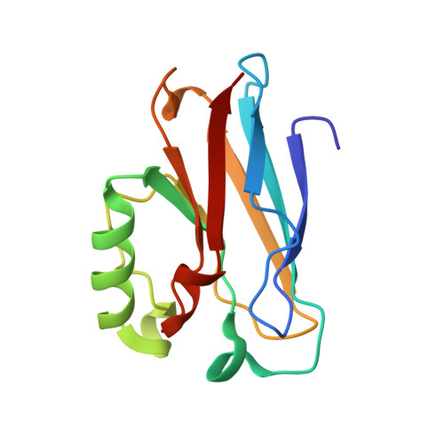
Crystal structure of the disulfide bond-deficient azurin mutant C3A/C26A: how important is the S-S bond for folding and stability?
Bonander, N., Leckner, J., Guo, H., Karlsson, B.G., Sjolin, L.(2000) Eur J Biochem 267: 4511-4519
- PubMed: 10880975 Search on PubMed
- DOI: https://doi.org/10.1046/j.1432-1033.2000.01501.x
- Primary Citation Related Structures:
1EZL - PubMed Abstract:
Azurin has a beta-barrel fold comprising eight beta-strands and one alpha helix. A disulfide bond between residues 3 and 26 connects the N-termini of beta strands beta1 and beta3. Three mutant proteins lacking the disulfide bond were constructed, C3A/C26A, C3A/C26I and a putative salt bridge (SB) in the C3A/S25R/C26A/K27R mutant. All three mutants exhibit spectroscopic properties similar to the wild-type protein. Furthermore, the crystal structure of the C3A/C26A mutant was determined at 2.0 A resolution and, in comparison to the wild-type protein, the only differences are found in the immediate proximity of the mutation. The mutants lose the 628 nm charge-transfer band at a temperature 10-22 degrees C lower than the wild-type protein. The folding of the zinc loaded C3A/C26A mutant was studied by guanidine hydrochloride (GdnHCl) induced denaturation monitored both by fluorescence and CD spectroscopy. The midpoint in the folding equilibrium, at 1.3 M GdnHCl, was observed using both CD and fluorescence spectroscopy. The free energy of folding determined from CD is -24.9 kJ.mol-1, a destabilization of approximately 20 kJ.mol-1 compared to the wild-type Zn2+-protein carrying an intact disulfide bond, indicating that the disulfide bond is important for giving azurin its stable structure. The C3A/C26I mutant is more stable and the SB mutant is less stable than C3A/C26A, both in terms of folding energy and thermal denaturation. The folding intermediate of the wild-type Zn2+-azurin is not observed for the disulfide-deficient C3A/C26A mutant. The rate of unfolding for the C3A/C26A mutant is similar to that of the wild-type protein, suggesting that the site of the mutation is not involved in an early unfolding reaction.
- Department of Biochemistry and Biophysics, Lundberg Institute, Göteborg University and Chalmers University of Technology, Sweden.
Organizational Affiliation: