1EZ1

STRUCTURE OF ESCHERICHIA COLI PURT-ENCODED GLYCINAMIDE RIBONUCLEOTIDE TRANSFORMYLASE COMPLEXED WITH MG, AMPPNP, AND GAR


Experimental Data Snapshot

  • Method: X-RAY DIFFRACTION
  • Resolution: 1.75 Å
  • R-Value Free: 0.254 
  • R-Value Work: 0.189 
  • R-Value Observed: 0.189 

wwPDB Validation   3D Report Full Report


Ligand Structure Quality Assessment 


This is version 1.4 of the entry. See complete history


Literature

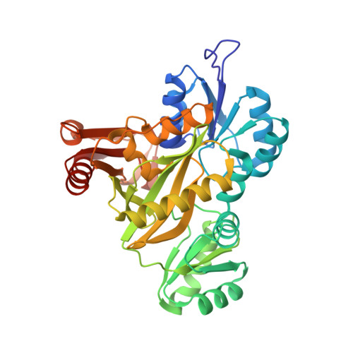
Molecular structure of Escherichia coli PurT-encoded glycinamide ribonucleotide transformylase.

Thoden, J.B.Firestine, S.Nixon, A.Benkovic, S.J.Holden, H.M.

(2000) Biochemistry 39: 8791-8802

  • DOI: https://doi.org/10.1021/bi000926j
  • Primary Citation of Related Structures:  
    1EYZ, 1EZ1

  • PubMed Abstract: 

    In Escherichia coli, the PurT-encoded glycinamide ribonucleotide transformylase, or PurT transformylase, catalyzes an alternative formylation of glycinamide ribonucleotide (GAR) in the de novo pathway for purine biosynthesis. On the basis of amino acid sequence analyses, it is known that the PurT transformylase belongs to the ATP-grasp superfamily of proteins. The common theme among members of this superfamily is a catalytic reaction mechanism that requires ATP and proceeds through an acyl phosphate intermediate. All of the enzymes belonging to the ATP-grasp superfamily are composed of three structural motifs, termed the A-, B-, and C-domains, and in each case, the ATP is wedged between the B- and C-domains. Here we describe two high-resolution X-ray crystallographic structures of PurT transformylase from E. coli: one form complexed with the nonhydrolyzable ATP analogue AMPPNP and the second with bound AMPPNP and GAR. The latter structure is of special significance because it represents the first ternary complex to be determined for a member of the ATP-grasp superfamily involved in purine biosynthesis and as such provides new information about the active site region involved in ribonucleotide binding. Specifically in PurT transformylase, the GAR substrate is anchored to the protein via Glu 82, Asp 286, Lys 355, Arg 362, and Arg 363. Key amino acid side chains involved in binding the AMPPNP to the enzyme include Arg 114, Lys 155, Glu 195, Glu 203, and Glu 267. Strikingly, the amino group of GAR that is formylated during the reaction lies at 2.8 A from one of the gamma-phosphoryl oxygens of the AMPPNP.


  • Organizational Affiliation

    Department of Biochemistry, University of Wisconsin, Madison, Wisconsin 53705, USA.


Macromolecules
Find similar proteins by:  (by identity cutoff)  |  3D Structure
Entity ID: 1
MoleculeChains Sequence LengthOrganismDetailsImage
PHOSPHORIBOSYLGLYCINAMIDE FORMYLTRANSFERASE 2
A, B
392Escherichia coliMutation(s): 0 
EC: 2.1.2
UniProt
Find proteins for P33221 (Escherichia coli (strain K12))
Explore P33221 
Go to UniProtKB:  P33221
Entity Groups  
Sequence Clusters30% Identity50% Identity70% Identity90% Identity95% Identity100% Identity
UniProt GroupP33221
Sequence Annotations
Expand
  • Reference Sequence
Small Molecules
Ligands 6 Unique
IDChains Name / Formula / InChI Key2D Diagram3D Interactions
ANP
Query on ANP

Download Ideal Coordinates CCD File 
G [auth A],
N [auth B]
PHOSPHOAMINOPHOSPHONIC ACID-ADENYLATE ESTER
C10 H17 N6 O12 P3
PVKSNHVPLWYQGJ-KQYNXXCUSA-N
GAR
Query on GAR

Download Ideal Coordinates CCD File 
H [auth A],
O [auth B]
GLYCINAMIDE RIBONUCLEOTIDE
C7 H13 N2 O8 P
OBQMLSFOUZUIOB-SHUUEZRQSA-L
MPO
Query on MPO

Download Ideal Coordinates CCD File 
I [auth A]3[N-MORPHOLINO]PROPANE SULFONIC ACID
C7 H15 N O4 S
DVLFYONBTKHTER-UHFFFAOYSA-N
ACT
Query on ACT

Download Ideal Coordinates CCD File 
E [auth A],
L [auth B]
ACETATE ION
C2 H3 O2
QTBSBXVTEAMEQO-UHFFFAOYSA-M
MG
Query on MG

Download Ideal Coordinates CCD File 
C [auth A],
D [auth A],
J [auth B],
K [auth B]
MAGNESIUM ION
Mg
JLVVSXFLKOJNIY-UHFFFAOYSA-N
NA
Query on NA

Download Ideal Coordinates CCD File 
F [auth A],
M [auth B]
SODIUM ION
Na
FKNQFGJONOIPTF-UHFFFAOYSA-N
Experimental Data & Validation

Experimental Data

  • Method: X-RAY DIFFRACTION
  • Resolution: 1.75 Å
  • R-Value Free: 0.254 
  • R-Value Work: 0.189 
  • R-Value Observed: 0.189 
  • Space Group: P 21 21 2
Unit Cell:
Length ( Å )Angle ( ˚ )
a = 61.8α = 90
b = 179.4β = 90
c = 75.3γ = 90
Software Package:
Software NamePurpose
TNTrefinement
FRAMBOdata collection
SAINTdata scaling
TNTphasing

Structure Validation

View Full Validation Report


Ligand Structure Quality Assessment 


Entry History 

Deposition Data

Revision History  (Full details and data files)

  • Version 1.0: 2000-08-02
    Type: Initial release
  • Version 1.1: 2008-04-27
    Changes: Version format compliance
  • Version 1.2: 2011-07-13
    Changes: Version format compliance
  • Version 1.3: 2017-10-04
    Changes: Refinement description
  • Version 1.4: 2024-02-07
    Changes: Data collection, Database references, Derived calculations