1ETU

STRUCTURAL DETAILS OF THE BINDING OF GUANOSINE DIPHOSPHATE TO ELONGATION FACTOR TU FROM E. COLI AS STUDIED BY X-RAY CRYSTALLOGRAPHY


Experimental Data Snapshot

  • Method: X-RAY DIFFRACTION
  • Resolution: 2.90 Å

wwPDB Validation   3D Report Full Report


This is version 1.2 of the entry. See complete history


Literature

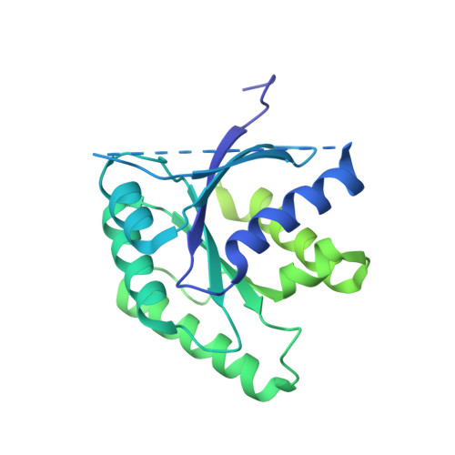
Structural details of the binding of guanosine diphosphate to elongation factor Tu from E. coli as studied by X-ray crystallography.

la Cour, T.F.Nyborg, J.Thirup, S.Clark, B.F.

(1985) EMBO J 4: 2385-2388

  • DOI: https://doi.org/10.1002/j.1460-2075.1985.tb03943.x
  • Primary Citation of Related Structures:  
    1ETU

  • PubMed Abstract: 

    Structural details of the guanosine diphosphate binding to a modified form of elongation factor Tu from Escherichia coli, resulting from X-ray crystallographic studies, are reported. The protein elements that take part in the nucleotide binding are located in four loops connecting beta-strands with alpha-helices. These loops correspond to regions in primary sequences which show a high degree of homology when compared with other prokaryotic and eukaryotic elongation factors and initiation factor 2.


Macromolecules
Find similar proteins by:  (by identity cutoff)  |  3D Structure
Entity ID: 1
MoleculeChains Sequence LengthOrganismDetailsImage
ELONGATION FACTOR TU393Escherichia coli K-12Mutation(s): 0 
UniProt
Find proteins for P0CE47 (Escherichia coli (strain K12))
Explore P0CE47 
Go to UniProtKB:  P0CE47
Entity Groups  
Sequence Clusters30% Identity50% Identity70% Identity90% Identity95% Identity100% Identity
UniProt GroupP0CE47
Sequence Annotations
Expand
  • Reference Sequence
Small Molecules
Ligands 2 Unique
IDChains Name / Formula / InChI Key2D Diagram3D Interactions
GDP
Query on GDP

Download Ideal Coordinates CCD File 
C [auth A]GUANOSINE-5'-DIPHOSPHATE
C10 H15 N5 O11 P2
QGWNDRXFNXRZMB-UUOKFMHZSA-N
MG
Query on MG

Download Ideal Coordinates CCD File 
B [auth A]MAGNESIUM ION
Mg
JLVVSXFLKOJNIY-UHFFFAOYSA-N
Experimental Data & Validation

Experimental Data

  • Method: X-RAY DIFFRACTION
  • Resolution: 2.90 Å
  • Space Group: C 2 2 21
Unit Cell:
Length ( Å )Angle ( ˚ )
a = 98.2α = 90
b = 100.8β = 90
c = 160.8γ = 90

Structure Validation

View Full Validation Report


Entry History 

Deposition Data

Revision History  (Full details and data files)

  • Version 1.0: 1988-07-16
    Type: Initial release
  • Version 1.1: 2008-03-24
    Changes: Version format compliance
  • Version 1.2: 2011-07-13
    Changes: Version format compliance