1ER8

THE ACTIVE SITE OF ASPARTIC PROTEINASES


Experimental Data Snapshot

  • Method: X-RAY DIFFRACTION
  • Resolution: 2.00 Å
  • R-Value Work: 0.170 

wwPDB Validation   3D Report Full Report


This is version 1.3 of the entry. See complete history


Literature

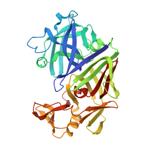
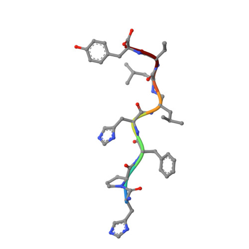
The Active Site of Aspartic Proteinases

Pearl, L.Blundell, T.

(1984) FEBS Lett 174: 96-101

  • DOI: https://doi.org/10.1016/0014-5793(84)81085-6
  • Primary Citation of Related Structures:  
    1ER8, 3ER3, 4APE, 4ER1, 4ER2

  • PubMed Abstract: 
  • The active site of the aspartic proteinase, endothiapepsin, has been defined by X-ray analysis and restrained least-squares refinement at 2.1 A resolution with a crystallographic agreement value of 0.16. The environments of the two catalytically important aspartyl groups are remarkably similar and the contributions of the NH2- and COOH-terminal domains to the catalytic centre are related by a local 2-fold axis ...

    The active site of the aspartic proteinase, endothiapepsin, has been defined by X-ray analysis and restrained least-squares refinement at 2.1 A resolution with a crystallographic agreement value of 0.16. The environments of the two catalytically important aspartyl groups are remarkably similar and the contributions of the NH2- and COOH-terminal domains to the catalytic centre are related by a local 2-fold axis. The carboxylates of the aspartyls share a hydrogen bond and have equivalent contacts to a bound water molecule or hydroxonium ion lying on the local diad. The main chains around 32 and 215 are connected by a novel interaction involving diad-related threonines. It is suggested that the two pKa values of the active site aspartyls arise from a structure not unlike that in maleic acid with a hydrogen-bonded intermediate species and a dicarboxylate characterised by electrostatic repulsions between the two negatively charged groups.


    Related Citations: 
    • Active Site of Acid Proteinases
      Blundell, T.L., Jones, H.B., Khan, G., Taylor, G., Sewell, T.S., Pearl, L.H., Wood, S.P.
      (1979) Proc Febs Meet 60: 281
    • The Three-Dimensional Structure of Acid Proteinases
      Blundell, T.L., Jenkins, J.A., Khan, G., Roychowdhury, P., Sewell, T., Tickle, I.J., Wood, E.A.
      (1979) Proc Febs Meet 52: 81
    • Four-Fold Structural Repeat in the Acid Proteases
      Blundell, T.L., Sewell, B.T., Mclachlan, A.D.
      (1979) Biochim Biophys Acta 580: 24
    • Structural Evidence for Gene Duplication in the Evolution of Acid Proteases
      Tang, J., James, M.N.G., Hsu, I.N., Jenkins, J.A., Blundell, T.L.
      (1978) Nature 271: 618
    • Homology Among Acid Proteases. Comparison of Crystal Structures at 3 Angstroms Resolution of Acid Proteases from Rhizopus Chinensis and Endothia Parasitica
      Subramanian, E., Swan, I.D.A., Liu, M., Davies, D.R., Jenkins, J.A., Tickle, I.J., Blundell, T.L.
      (1977) Proc Natl Acad Sci U S A 74: 556
    • X-Ray Analysis and Circular Dichroism of the Acid Protease from Endothia Parasitica and Chymosin
      Jenkins, J., Tickle, I., Sewell, T., Ungaretti, L., Wollmer, A., Blundell, T.
      (1977) Adv Exp Med Biol 95: 43

    Organizational Affiliation

    Department of Biophysics, All India Institute of Medical Sciences, New Delhi, India.


Macromolecules
Find similar proteins by:  (by identity cutoff)  |  3D Structure
Entity ID: 1
MoleculeChainsSequence LengthOrganismDetailsImage
EndothiapepsinA [auth E]330Cryphonectria parasiticaMutation(s): 0 
Gene Names: EAPAEPN-1
EC: 3.4.23.22
UniProt
Find proteins for P11838 (Cryphonectria parasitica)
Explore P11838 
Go to UniProtKB:  P11838
Entity Groups  
Sequence Clusters30% Identity50% Identity70% Identity90% Identity95% Identity100% Identity
UniProt GroupP11838
Protein Feature View
Expand
  • Reference Sequence

Find similar proteins by:  Sequence   |   3D Structure  

Entity ID: 2
MoleculeChainsSequence LengthOrganismDetailsImage
H-77B [auth I]8N/AMutation(s): 0 
UniProt
Find proteins for P01016 (Equus caballus)
Explore P01016 
Go to UniProtKB:  P01016
Entity Groups  
UniProt GroupP01016
Protein Feature View
Expand
  • Reference Sequence
Experimental Data & Validation

Experimental Data

  • Method: X-RAY DIFFRACTION
  • Resolution: 2.00 Å
  • R-Value Work: 0.170 
  • Space Group: P 1 21 1
Unit Cell:
Length ( Å )Angle ( ˚ )
a = 43.2α = 90
b = 75.7β = 97.1
c = 42.9γ = 90

Structure Validation

View Full Validation Report


Entry History 

Deposition Data

Revision History  (Full details and data files)

  • Version 1.0: 1991-10-15
    Type: Initial release
  • Version 1.1: 2008-03-03
    Changes: Version format compliance
  • Version 1.2: 2011-07-13
    Changes: Version format compliance
  • Version 1.3: 2017-11-29
    Changes: Derived calculations, Other