1EF0 | pdb_00001ef0

CRYSTAL STRUCTURE OF PI-SCEI MINIPRECURSOR


Experimental Data Snapshot

  • Method: X-RAY DIFFRACTION
  • Resolution: 2.10 Å
  • R-Value Free: 
    0.281 (Depositor), 0.275 (DCC) 
  • R-Value Work: 
    0.231 (Depositor), 0.226 (DCC) 
  • R-Value Observed: 
    0.231 (Depositor) 

wwPDB Validation 3D Report Full Report

Validation slider image for 1EF0

This is version 1.5 of the entry. See complete history

Literature

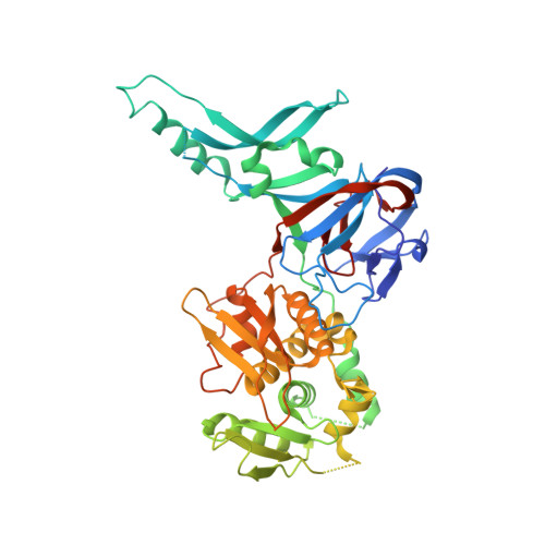
Structural insights into the protein splicing mechanism of PI-SceI.

Poland, B.W.Xu, M.Q.Quiocho, F.A.

(2000) J Biological Chem 275: 16408-16413

  • DOI: https://doi.org/10.1074/jbc.275.22.16408
  • Primary Citation Related Structures: 
    1EF0

  • PubMed Abstract: 

    PI-SceI is a member of a class of proteins (inteins) that excise themselves from a precursor protein and in the process ligate the flanking protein sequences (exteins). We report here the 2.1-A resolution crystal structure of a PI-SceI miniprecursor (VMA29) containing 10 N-terminal extein residues and 4 C-terminal extein residues. Mutations at the N- and C-terminal splicing junctions, blocking in vivo protein splicing, allowed the miniprecursor to be purified and crystallized. The structure reveals both the N- and C-terminal scissile peptide bonds to be in distorted trans conformations (tau approximately 100 degrees ). Modeling of the wild-type PI-SceI based on the VMA29 structure indicates a large conformational change (movement of >9 A) must occur to allow transesterification to be completed. A zinc atom was discovered at the C-terminal splicing junction. Residues Cys(455), His(453), and Glu(80) along with a water molecule (Wat(53)) chelate the zinc atom. The crystal structure of VMA29 has captured the intein in its pre-spliced state.


  • Organizational Affiliation
    • Howard Hughes Medical Institute and Department of Biochemistry, Baylor College of Medicine, Houston, Texas 77030, USA.

Macromolecule Content 

  • Total Structure Weight: 103.74 kDa 
  • Atom Count: 6,887 
  • Modeled Residue Count: 819 
  • Deposited Residue Count: 924 
  • Unique protein chains: 1

Macromolecules

Find similar proteins by:|  3D Structure
Entity ID: 1
MoleculeChains  Sequence LengthOrganismDetailsImage
PI-SCEI ENDONUCLEASE
A, B
462Saccharomyces cerevisiaeMutation(s): 2 
EC: 7.1.2.2
UniProt
Find proteins for P17255 (Saccharomyces cerevisiae (strain ATCC 204508 / S288c))
Explore P17255 
Go to UniProtKB:  P17255
Entity Groups
Sequence Clusters30% Identity50% Identity70% Identity90% Identity95% Identity100% Identity
UniProt GroupP17255
Sequence Annotations
Expand
Reference Sequence

Experimental Data & Validation

Experimental Data

  • Method: X-RAY DIFFRACTION
  • Resolution: 2.10 Å
  • R-Value Free:  0.281 (Depositor), 0.275 (DCC) 
  • R-Value Work:  0.231 (Depositor), 0.226 (DCC) 
  • R-Value Observed: 0.231 (Depositor) 
Space Group: P 1 21 1
Unit Cell:
Length ( Å )Angle ( ˚ )
a = 59.003α = 90
b = 101.683β = 93.51
c = 86.765γ = 90
Software Package:
Software NamePurpose
CNSrefinement
XTALVIEWrefinement
DENZOdata reduction
SCALEPACKdata scaling

Structure Validation

View Full Validation Report


Entry History 

Deposition Data

Revision History  (Full details and data files)

  • Version 1.0: 2000-06-07
    Type: Initial release
  • Version 1.1: 2008-04-27
    Changes: Version format compliance
  • Version 1.2: 2011-07-13
    Changes: Version format compliance
  • Version 1.3: 2019-07-24
    Changes: Data collection, Refinement description
  • Version 1.4: 2021-11-03
    Changes: Database references, Derived calculations
  • Version 1.5: 2024-02-07
    Changes: Data collection