Sequence-specific RNA binding by a Nova KH domain: implications for paraneoplastic disease and the fragile X syndrome.
Lewis, H.A., Musunuru, K., Jensen, K.B., Edo, C., Chen, H., Darnell, R.B., Burley, S.K.(2000) Cell 100: 323-332
- PubMed: 10676814 Search on PubMed
- DOI: https://doi.org/10.1016/s0092-8674(00)80668-6
- Primary Citation Related Structures:
1EC6 - PubMed Abstract:
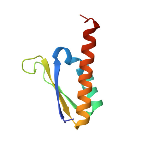
The structure of a Nova protein K homology (KH) domain recognizing single-stranded RNA has been determined at 2.4 A resolution. Mammalian Nova antigens (1 and 2) constitute an important family of regulators of RNA metabolism in neurons, first identified using sera from cancer patients with the autoimmune disorder paraneoplastic opsoclonus-myoclonus ataxia (POMA). The structure of the third KH domain (KH3) of Nova-2 bound to a stem loop RNA resembles a molecular vise, with 5'-Ura-Cyt-Ade-Cyt-3' pinioned between an invariant Gly-X-X-Gly motif and the variable loop. Tetranucleotide recognition is supported by an aliphatic alpha helix/beta sheet RNA-binding platform, which mimics 5'-Ura-Gua-3' by making Watson-Crick-like hydrogen bonds with 5'-Cyt-Ade-3'. Sequence conservation suggests that fragile X mental retardation results from perturbation of RNA binding by the FMR1 protein.
- Laboratories of Molecular Biophysics, The Rockefeller University, New York, New York 10021, USA.
Organizational Affiliation: