1EAY

CHEY-BINDING (P2) DOMAIN OF CHEA IN COMPLEX WITH CHEY FROM ESCHERICHIA COLI


Experimental Data Snapshot

  • Method: X-RAY DIFFRACTION
  • Resolution: 2.00 Å
  • R-Value Work: 0.217 

wwPDB Validation   3D Report Full Report


This is version 1.3 of the entry. See complete history


Literature

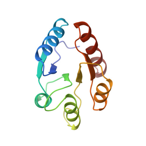
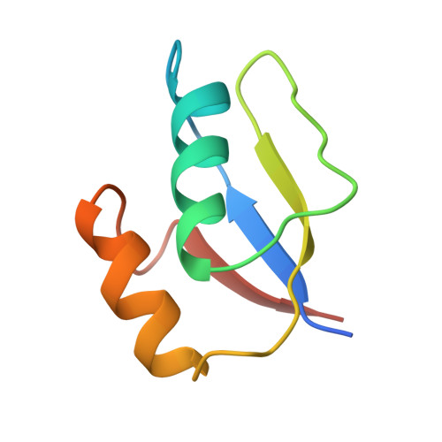
Two binding modes reveal flexibility in kinase/response regulator interactions in the bacterial chemotaxis pathway.

McEvoy, M.M.Hausrath, A.C.Randolph, G.B.Remington, S.J.Dahlquist, F.W.

(1998) Proc Natl Acad Sci U S A 95: 7333-7338

  • DOI: https://doi.org/10.1073/pnas.95.13.7333
  • Primary Citation of Related Structures:  
    1EAY

  • PubMed Abstract: 

    The crystal structure at 2.0-A resolution of the complex of the Escherichia coli chemotaxis response regulator CheY and the phosphoacceptor-binding domain (P2) of the kinase CheA is presented. The binding interface involves the fourth and fifth helices and fifth beta-strand of CheY and both helices of P2. Surprisingly, the two heterodimers in the asymmetric unit have two different binding modes involving the same interface, suggesting some flexibility in the binding regions. Significant conformational changes have occurred in CheY compared with previously determined unbound structures. The active site of CheY is exposed by the binding of the kinase domain, possibly to enhance phosphotransfer from CheA to CheY. The conformational changes upon complex formation as well as the observation that there are two different binding modes suggest that the plasticity of CheY is an essential feature of response regulator function.


  • Organizational Affiliation

    Institute of Molecular Biology, University of Oregon, Eugene, OR 97403, USA.


Macromolecules
Find similar proteins by:  (by identity cutoff)  |  3D Structure
Entity ID: 1
MoleculeChains Sequence LengthOrganismDetailsImage
CHEY
A, B
128Escherichia coliMutation(s): 0 
UniProt
Find proteins for P0AE67 (Escherichia coli (strain K12))
Explore P0AE67 
Go to UniProtKB:  P0AE67
Entity Groups  
Sequence Clusters30% Identity50% Identity70% Identity90% Identity95% Identity100% Identity
UniProt GroupP0AE67
Sequence Annotations
Expand
  • Reference Sequence
Find similar proteins by:  (by identity cutoff)  |  3D Structure
Entity ID: 2
MoleculeChains Sequence LengthOrganismDetailsImage
CHEA
C, D
74Escherichia coliMutation(s): 0 
EC: 2.7.3
UniProt
Find proteins for P07363 (Escherichia coli (strain K12))
Explore P07363 
Go to UniProtKB:  P07363
Entity Groups  
Sequence Clusters30% Identity50% Identity70% Identity90% Identity95% Identity100% Identity
UniProt GroupP07363
Sequence Annotations
Expand
  • Reference Sequence
Experimental Data & Validation

Experimental Data

  • Method: X-RAY DIFFRACTION
  • Resolution: 2.00 Å
  • R-Value Work: 0.217 
  • Space Group: P 21 21 21
Unit Cell:
Length ( Å )Angle ( ˚ )
a = 54.5α = 90
b = 64.2β = 90
c = 158γ = 90
Software Package:
Software NamePurpose
PROTEINmodel building
SIGMAAmodel building
RSSmodel building
TNTrefinement
DENZOdata reduction
SCALEPACKdata scaling
PROTEINphasing
SIGMAAphasing
RSSphasing

Structure Validation

View Full Validation Report


Entry History 

Deposition Data

Revision History  (Full details and data files)

  • Version 1.0: 1998-07-15
    Type: Initial release
  • Version 1.1: 2008-03-24
    Changes: Version format compliance
  • Version 1.2: 2011-07-13
    Changes: Version format compliance
  • Version 1.3: 2023-08-09
    Changes: Database references, Other, Refinement description