1E8U

Structure of the multifunctional paramyxovirus hemagglutinin-neuraminidase


Experimental Data Snapshot

  • Method: X-RAY DIFFRACTION
  • Resolution: 2.00 Å
  • R-Value Free: 0.267 
  • R-Value Work: 0.232 
  • R-Value Observed: 0.232 

wwPDB Validation   3D Report Full Report


This is version 1.5 of the entry. See complete history


Literature

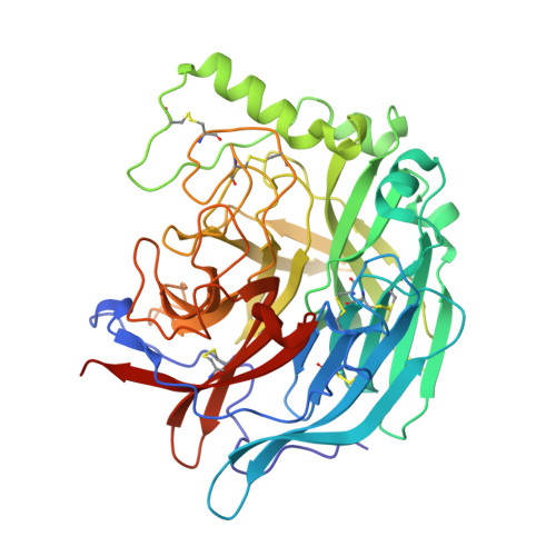
Crystal Structure of the Multifunctional Paramyxovirus Hemagglutinin-Neuraminidase

Crennell, S.Takimoto, T.Portner, A.Taylor, G.

(2000) Nat Struct Biol 7: 1068

  • DOI: https://doi.org/10.1038/81002
  • Primary Citation of Related Structures:  
    1E8T, 1E8U, 1E8V

  • PubMed Abstract: 

    Paramyxoviruses are the main cause of respiratory disease in children. One of two viral surface glycoproteins, the hemagglutinin-neuraminidase (HN), has several functions in addition to being the major surface antigen that induces neutralizing antibodies. Here we present the crystal structures of Newcastle disease virus HN alone and in complex with either an inhibitor or with the beta-anomer of sialic acid. The inhibitor complex reveals a typical neuraminidase active site within a beta-propeller fold. Comparison of the structures of the two complexes reveal differences in the active site, suggesting that the catalytic site is activated by a conformational switch. This site may provide both sialic acid binding and hydrolysis functions since there is no evidence for a second sialic acid binding site in HN. Evidence for a single site with dual functions is examined and supported by mutagenesis studies. The structure provides the basis for the structure-based design of inhibitors for a range of paramyxovirus-induced diseases.


  • Organizational Affiliation

    Department of Biology and Biochemistry, University of Bath, Bath BA2 7AY, UK.


Macromolecules
Find similar proteins by:  (by identity cutoff)  |  3D Structure
Entity ID: 1
MoleculeChains Sequence LengthOrganismDetailsImage
HEMAGGLUTININ-NEURAMINIDASE
A, B
454avian paramyxovirus 1Mutation(s): 0 
EC: 3.2.1.18
UniProt
Find proteins for Q9Q2W5 (Newcastle disease virus (strain Kansas))
Explore Q9Q2W5 
Go to UniProtKB:  Q9Q2W5
Entity Groups  
Sequence Clusters30% Identity50% Identity70% Identity90% Identity95% Identity100% Identity
UniProt GroupQ9Q2W5
Sequence Annotations
Expand
  • Reference Sequence
Experimental Data & Validation

Experimental Data

  • Method: X-RAY DIFFRACTION
  • Resolution: 2.00 Å
  • R-Value Free: 0.267 
  • R-Value Work: 0.232 
  • R-Value Observed: 0.232 
  • Space Group: P 21 21 21
Unit Cell:
Length ( Å )Angle ( ˚ )
a = 72.56α = 90
b = 78.49β = 90
c = 199.93γ = 90
Software Package:
Software NamePurpose
CNSrefinement

Structure Validation

View Full Validation Report


Entry History 

Deposition Data

Revision History  (Full details and data files)

  • Version 1.0: 2001-04-03
    Type: Initial release
  • Version 1.1: 2011-11-09
    Changes: Advisory, Atomic model, Derived calculations, Non-polymer description, Other, Refinement description, Structure summary, Version format compliance
  • Version 1.2: 2018-05-02
    Changes: Data collection
  • Version 1.3: 2019-07-10
    Changes: Data collection, Derived calculations
  • Version 1.4: 2019-07-24
    Changes: Data collection
  • Version 1.5: 2020-07-29
    Type: Remediation
    Reason: Carbohydrate remediation
    Changes: Advisory, Data collection, Derived calculations, Structure summary