1E29 | pdb_00001e29

PSII associated cytochrome C549 from Synechocystis sp.


Experimental Data Snapshot

  • Method: X-RAY DIFFRACTION
  • Resolution: 1.21 Å
  • R-Value Free: 
    0.212 (Depositor) 
  • R-Value Work: 
    0.151 (DCC) 
  • R-Value Observed: 
    0.149 (Depositor) 

wwPDB Validation   3D Report Full Report


Ligand Structure Quality Assessment 

Created with Raphaël 2.3.0Worse 01 BetterLigand structure goodness of fit to experimental dataBest fitted HECClick on this verticalbar to view details

This is version 1.7 of the entry. See complete history


Literature

Crystal Structure of Low-Potential Cytochrome C549 from Synechocystis Sp. Pcc 6803 at 1.21A Resolution

Frazao, C.Enguita, F.J.Coelho, R.Sheldrick, G.M.Navarro, J.A.Hervas, M.De La Rosa, M.A.Carrondo, M.A.

(2001) J Biol Inorg Chem 6: 324

  • DOI: https://doi.org/10.1007/s007750100208
  • Primary Citation of Related Structures:  
    1E29

  • PubMed Abstract: 

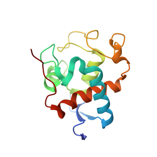
    The crystal structure of low-potential cytochrome c549, an extrinsic component of the photosystem II (PS II) from Synechocystis sp. PCC 6803, was obtained directly from single-wavelength 1.21 A resolution diffraction data. This is the first monodomain bis-histidinyl monoheme cytochrome c to be structurally characterized. The extended N-terminal region of c549 builds up a two-strand antiparallel beta-sheet in a hairpin motif, which extends through two molecules owing to crystal packing. Both peptide termini are involved in crystal contacts, which may explain their protrusion out of the globular fold. The C-terminus is preceded by a 9 A-long hydrophobic finger extending from a positively charged base and could be involved in PSII interactions, as well as a protruding negative patch built by a set of conserved acidic residues among c549 sequences.


  • Organizational Affiliation

    Instituto de Tecnologia Química e Biológica, Universidade Nova de Lisboa, 2781-901 Oeiras, Portugal.


Macromolecules
Find similar proteins by:  (by identity cutoff)  |  3D Structure
Entity ID: 1
MoleculeChains Sequence LengthOrganismDetailsImage
CYTOCHROME C549135Synechocystis sp.Mutation(s): 0 
UniProt
Find proteins for Q55013 (Synechocystis sp. (strain ATCC 27184 / PCC 6803 / Kazusa))
Explore Q55013 
Go to UniProtKB:  Q55013
Entity Groups  
Sequence Clusters30% Identity50% Identity70% Identity90% Identity95% Identity100% Identity
UniProt GroupQ55013
Sequence Annotations
Expand
  • Reference Sequence
Experimental Data & Validation

Experimental Data

  • Method: X-RAY DIFFRACTION
  • Resolution: 1.21 Å
  • R-Value Free:  0.212 (Depositor) 
  • R-Value Work:  0.151 (DCC) 
  • R-Value Observed: 0.149 (Depositor) 
Space Group: C 2 2 21
Unit Cell:
Length ( Å )Angle ( ˚ )
a = 34.22α = 90
b = 86.86β = 90
c = 97.66γ = 90
Software Package:
Software NamePurpose
SHELXL-97refinement
DENZOdata reduction
SCALEPACKdata scaling
SHELXDphasing

Structure Validation

View Full Validation Report


Ligand Structure Quality Assessment 

Created with Raphaël 2.3.0Worse 01 BetterLigand structure goodness of fit to experimental dataBest fitted HECClick on this verticalbar to view details

Entry History 

Deposition Data

Revision History  (Full details and data files)

  • Version 1.0: 2001-05-04
    Type: Initial release
  • Version 1.1: 2011-05-08
    Changes: Version format compliance
  • Version 1.2: 2011-07-13
    Changes: Version format compliance
  • Version 1.3: 2017-07-12
    Changes: Derived calculations
  • Version 1.4: 2019-05-08
    Changes: Data collection, Experimental preparation
  • Version 1.5: 2019-05-22
    Changes: Data collection, Refinement description
  • Version 1.6: 2019-07-24
    Changes: Data collection
  • Version 1.7: 2024-11-06
    Changes: Data collection, Database references, Derived calculations, Other, Structure summary