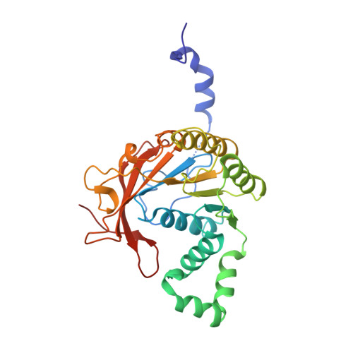
1E0J

gp4d helicase from phage T7 ADPNP complex


Experimental Data Snapshot

  • Method: X-RAY DIFFRACTION
  • Resolution: 3.00 Å
  • R-Value Free: 0.304 
  • R-Value Work: 0.253 
  • R-Value Observed: 0.253 

wwPDB Validation   3D Report Full Report


Ligand Structure Quality Assessment 


This is version 1.3 of the entry. See complete history


Literature

Crystal Structure of T7 Gene 4 Ring Helicase Indicates a Mechanism for Sequential Hydrolysis of Nucleotides

Singleton, M.R.Sawaya, M.R.Ellenberger, T.Wigley, D.B.

(2000) Cell 101: 589

  • DOI: https://doi.org/10.1016/s0092-8674(00)80871-5
  • Primary Citation of Related Structures:  
    1E0J, 1E0K

  • PubMed Abstract: 

    We have determined the crystal structure of an active, hexameric fragment of the gene 4 helicase from bacteriophage T7. The structure reveals how subunit contacts stabilize the hexamer. Deviation from expected six-fold symmetry of the hexamer indicates that the structure is of an intermediate on the catalytic pathway. The structural consequences of the asymmetry suggest a "binding change" mechanism to explain how cooperative binding and hydrolysis of nucleotides are coupled to conformational changes in the ring that most likely accompany duplex unwinding. The structure of a complex with a nonhydrolyzable ATP analog provides additional evidence for this hypothesis, with only four of the six possible nucleotide binding sites being occupied in this conformation of the hexamer. This model suggests a mechanism for DNA translocation.


  • Organizational Affiliation

    Sir William Dunn School of Pathology, University of Oxford, United Kingdom.


Macromolecules
Find similar proteins by:  (by identity cutoff)  |  3D Structure
Entity ID: 1
MoleculeChains Sequence LengthOrganismDetailsImage
DNA HELICASE
A, B, C, D, E
A, B, C, D, E, F
289Escherichia phage T7Mutation(s): 0 
Gene Names: GENE 4
UniProt
Find proteins for P03692 (Escherichia phage T7)
Explore P03692 
Go to UniProtKB:  P03692
Entity Groups  
Sequence Clusters30% Identity50% Identity70% Identity90% Identity95% Identity100% Identity
UniProt GroupP03692
Sequence Annotations
Expand
  • Reference Sequence
Experimental Data & Validation

Experimental Data

  • Method: X-RAY DIFFRACTION
  • Resolution: 3.00 Å
  • R-Value Free: 0.304 
  • R-Value Work: 0.253 
  • R-Value Observed: 0.253 
  • Space Group: P 41 21 2
Unit Cell:
Length ( Å )Angle ( ˚ )
a = 119.18α = 90
b = 119.18β = 90
c = 283.42γ = 90
Software Package:
Software NamePurpose
CNSrefinement
DENZOdata reduction
SCALEPACKdata scaling
CNSphasing

Structure Validation

View Full Validation Report


Ligand Structure Quality Assessment 


Entry History 

Deposition Data

Revision History  (Full details and data files)

  • Version 1.0: 2000-06-09
    Type: Initial release
  • Version 1.1: 2011-05-08
    Changes: Version format compliance
  • Version 1.2: 2011-07-13
    Changes: Version format compliance
  • Version 1.3: 2024-05-08
    Changes: Data collection, Database references, Derived calculations, Other