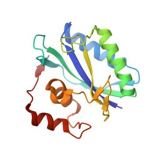
2.0A X-Ray Structure of the Ternary Complex of 7,8-Dihydro-6-Hydroxymethylpterinpyrophosphokinase from Escherichia Coli with ATP and a Substrate Analogue
Stammers, D.K., Achari, A., Somers, D.O., Bryant, P.K., Rosemond, J., Scott, D.L., Champness, J.N.(1999) FEBS Lett 456: 49
- PubMed: 10452528
- DOI: https://doi.org/10.1016/s0014-5793(99)00860-1
- Primary Citation of Related Structures:
1DY3 - PubMed Abstract:
The X-ray crystal structure of 7,8-dihydro-6-hydroxymethylpterinpyrophosphokinase (PPPK) in a ternary complex with ATP and a pterin analogue has been solved to 2.0 A resolution, giving, for the first time, detailed information of the PPPK/ATP intermolecular interactions and the accompanying conformational change. The first 100 residues of the 158 residue peptide contain a betaalpha betabeta alphabeta motif present in several other proteins including nucleoside diphosphate kinase. Comparative sequence examination of a wide range of prokaryotic and lower eukaryotic species confirms the conservation of the PPPK active site, indicating the value of this de novo folate biosynthesis pathway enzyme as a potential target for the development of novel broad-spectrum anti-infective agents.
- Glaxo Wellcome R&D, Medicines Research Centre, Stevenage, UK.
Organizational Affiliation:


