The Structure of the N-Terminal Actin-Binding Domain of Human Dystrophin and How Mutations in This Domain May Cause Duchenne or Becker Muscular Dystrophy
Norwood, F.L., Sutherland-Smith, A.J., Keep, N.H., Kendrick-Jones, J.(2000) Structure 8: 481
- PubMed: 10801490 Search on PubMed
- DOI: https://doi.org/10.1016/s0969-2126(00)00132-5
- Primary Citation Related Structures:
1DXX - PubMed Abstract:
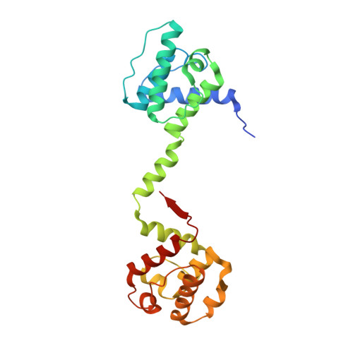
Dystrophin is an essential component of skeletal muscle cells. Its N-terminal domain binds to F-actin and its C terminus binds to the dystrophin-associated glycoprotein (DAG) complex in the membrane. Dystrophin is therefore thought to serve as a link from the actin-based cytoskeleton of the muscle cell through the plasma membrane to the extracellular matrix. Pathogenic mutations in dystrophin result in Duchenne or Becker muscular dystrophy. The crystal structure of the dystrophin actin-binding domain (ABD) has been determined at 2.6 A resolution. The structure is an antiparallel dimer of two ABDs each comprising two calponin homology domains (CH1 and CH2) that are linked by a central alpha helix. The CH domains are both alpha-helical globular folds. Comparisons with the structures of utrophin and fimbrin ABDs reveal that the conformations of the individual CH domains are very similar to those of dystrophin but that the arrangement of the two CH domains within the ABD is altered. The dystrophin dimer reveals a change of 72 degrees in the orientation of one pair of CH1 and CH2 domains (from different monomers) relative to the other pair when compared with the utrophin dimer. The dystrophin monomer is more elongated than the fimbrin ABD. The dystrophin ABD structure reveals a previously uncharacterised arrangement of the CH domains within the ABD. This observation has implications for the mechanism of actin binding by dystrophin and related proteins. Examining the position of three pathogenic missense mutations within the structure suggests that they exert their effects through misfolding of the ABD, rather than through disruption of the binding to F-actin.
- Structural Studies Division, Medical Research Council Laboratory of Molecular Biology, Cambridge, CB2 2QH, UK.
Organizational Affiliation: