1DOZ

CRYSTAL STRUCTURE OF FERROCHELATASE


Experimental Data Snapshot

  • Method: X-RAY DIFFRACTION
  • Resolution: 1.80 Å
  • R-Value Free: 0.216 
  • R-Value Work: 0.183 
  • R-Value Observed: 0.183 

wwPDB Validation   3D Report Full Report


This is version 1.4 of the entry. See complete history


Literature

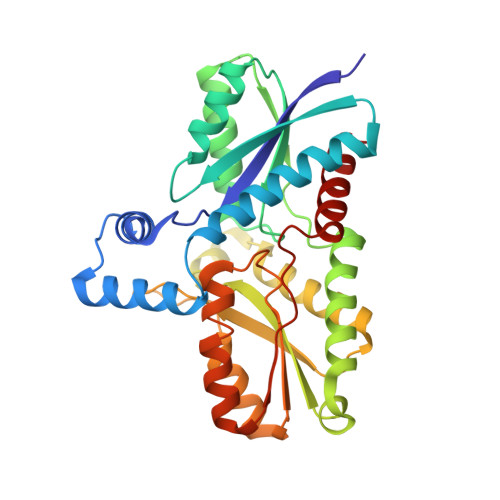
Structural and mechanistic basis of porphyrin metallation by ferrochelatase.

Lecerof, D.Fodje, M.Hansson, A.Hansson, M.Al-Karadaghi, S.

(2000) J Mol Biol 297: 221-232

  • DOI: 10.1006/jmbi.2000.3569
  • Primary Citation of Related Structures:  
    1C1H, 1C9E, 1DOZ

  • PubMed Abstract: 
  • Ferrochelatase, the enzyme catalyzing metallation of protoporphyrin IX at the terminal step of heme biosynthesis, was co-crystallized with an isomer mixture of the potent inhibitor N-methylmesoporphyrin (N-MeMP). The X-ray structure revealed the active site of the enzyme, to which only one of the isomers was bound, and for the first time allowed characterization of the mode of porphyrin macrocycle distortion by ferrochelatase ...

    Ferrochelatase, the enzyme catalyzing metallation of protoporphyrin IX at the terminal step of heme biosynthesis, was co-crystallized with an isomer mixture of the potent inhibitor N-methylmesoporphyrin (N-MeMP). The X-ray structure revealed the active site of the enzyme, to which only one of the isomers was bound, and for the first time allowed characterization of the mode of porphyrin macrocycle distortion by ferrochelatase. Crystallization of ferrochelatase and N-MeMP in the presence of Cu(2+) leads to metallation and demethylation of N-MeMP. A mechanism of porphyrin distortion is proposed, which assumes that the enzyme holds pyrrole rings B, C and D in a vice-like grip and forces a 36 degrees tilt on ring A.


    Organizational Affiliation

    Department of Molecular Biophysics, Lund University, Sweden.


Macromolecules
Find similar proteins by:  (by identity cutoff)  |  3D Structure
Entity ID: 1
MoleculeChainsSequence LengthOrganismDetailsImage
FERROCHELATASEA309Bacillus subtilisMutation(s): 0 
EC: 4.99.1.1 (PDB Primary Data), 4.99.1.9 (UniProt)
UniProt
Find proteins for P32396 (Bacillus subtilis (strain 168))
Explore P32396 
Go to UniProtKB:  P32396
Entity Groups  
Sequence Clusters30% Identity50% Identity70% Identity90% Identity95% Identity100% Identity
UniProt GroupP32396
Protein Feature View
Expand
  • Reference Sequence
Small Molecules
Ligands 1 Unique
IDChainsName / Formula / InChI Key2D Diagram3D Interactions
MG
Query on MG

Download Ideal Coordinates CCD File 
B [auth A],
C [auth A],
D [auth A]
MAGNESIUM ION
Mg
JLVVSXFLKOJNIY-UHFFFAOYSA-N
 Ligand Interaction
Experimental Data & Validation

Experimental Data

  • Method: X-RAY DIFFRACTION
  • Resolution: 1.80 Å
  • R-Value Free: 0.216 
  • R-Value Work: 0.183 
  • R-Value Observed: 0.183 
  • Space Group: P 21 21 21
Unit Cell:
Length ( Å )Angle ( ˚ )
a = 48.51α = 90
b = 49.97β = 90
c = 119.24γ = 90
Software Package:
Software NamePurpose
CNSrefinement
MAR345data collection
SCALEPACKdata scaling
CNSphasing

Structure Validation

View Full Validation Report


Entry History 

Deposition Data

Revision History  (Full details and data files)

  • Version 1.0: 2000-01-12
    Type: Initial release
  • Version 1.1: 2008-04-27
    Changes: Version format compliance
  • Version 1.2: 2011-07-13
    Changes: Version format compliance
  • Version 1.3: 2014-11-12
    Changes: Structure summary
  • Version 1.4: 2017-10-04
    Changes: Data collection, Refinement description