The structural basis for molecular recognition by the vitamin B 12 RNA aptamer.
Sussman, D., Nix, J.C., Wilson, C.(2000) Nat Struct Biol 7: 53-57
- PubMed: 10625428 Search on PubMed
- DOI: https://doi.org/10.1038/71253
- Primary Citation Related Structures:
1DDY - PubMed Abstract:
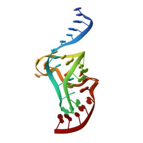
Previous solution structures of ligand-binding RNA aptamers have shown that molecular recognition is achieved by the folding of an initially unstructured RNA around its cognate ligand, coupling the processes of RNA folding and binding. The 3 A crystal structure of the cyanocobalamin (vitamin B12) aptamer reported here suggests a different approach to molecular recognition in which elements of RNA secondary structure combine to create a solvent-accessible docking surface for a large, complex ligand. Central to this structure is a locally folding RNA triplex, stabilized by a novel three-stranded zipper. Perpendicular stacking of a duplex on this triplex creates a cleft that functions as the vitamin B12 binding site. Complementary packing of hydrophobic surfaces, direct hydrogen bonding and dipolar interactions between the ligand and the RNA appear to contribute to binding. The nature of the interactions that stabilize complex formation and the possible uncoupling of folding and binding for this RNA suggest a strong mechanistic similarity to typical protein-ligand complexes.
- Department of Biology and Center for the Molecular Biology of RNA, Sinsheimer Laboratories, University of California, Santa Cruz, California 95064, USA. sussman@biology.ucsc.edu
Organizational Affiliation: