Mechanism of an ATP-dependent carboxylase, dethiobiotin synthetase, based on crystallographic studies of complexes with substrates and a reaction intermediate.
Huang, W., Jia, J., Gibson, K.J., Taylor, W.S., Rendina, A.R., Schneider, G., Lindqvist, Y.(1995) Biochemistry 34: 10985-10995
- PubMed: 7669756 Search on PubMed
- DOI: https://doi.org/10.1021/bi00035a004
- Primary Citation Related Structures:
1DAD, 1DAE, 1DAF, 1DAG, 1DAH, 1DAI - PubMed Abstract:
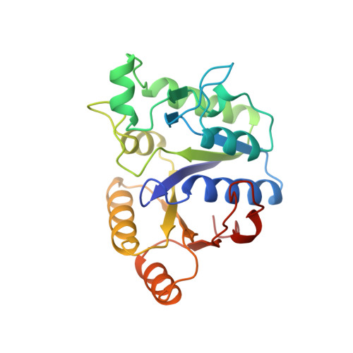
The crystal structures of six complexes of homodimeric Escherichia coli dethiobiotin synthetase with a variety of substrates, substrate analogs, and products have been determined to high resolution. These include (1) the binary complex of dethiobiotin synthetase and the N7-carbamate of 7,8-diaminononanoic acid, (2) the binary complex of enzyme and the alternate substrate, 3-(1-aminoethyl)-nonanedioic acid, (3) the binary complex of enzyme with the product ADP, (4) the quaternary complex of enzyme, ADP, the N7-carbamate of 7,8-diaminononanoic acid, and Ca2+, (5) the ternary complex of enzyme, the ATP analog adenylyl (beta, gamma-methylene)diphosphonate, and the N7-carbamate of 7,8-diaminononanoic acid, and (6) the quaternary complex of enzyme, the ATP analog adenylyl (beta, gamma-methylene)diphosphonate, 7,8-diaminononanoic acid, and Mn2+. One molecule of each substrate binds to one monomer of the enzyme. ADP and the ATP analogue bind to the classical mononucleotide binding fold with the phosphate groups close to the phosphate binding loop Gly8--Thr16 between beta-strand beta 1 and the N-terminus of alpha-helix alpha 1. The adenine ring is bound in a pocket between beta-strands beta 6 and beta 7. In the quaternary complex with Mn2+, the metal binding site is found in the vicinity of the beta- and gamma-phosphate groups. Two oxygen atoms from the phosphates and oxygen atoms from the side chains of Asp54, Thr16, and Glu115 are ligands to the Mn2+ ion in the quaternary complex. In the complex with ADP and the N7-carbamate of 7,8-diaminononanoic acid prepared in the presence of Ca2+ ions, a different metal binding site is found. The Ca2+ ion is coordinated to an oxygen atom of the alpha-phosphate group of the nucleotide, the side chain of Asp54, and solvent molecules. The 7,8-diaminononanoic acid substrate molecule interacts with residues from both subunits, making the dimer the minimal functional unit. The diamino group binds between the loops after beta 2 and beta 4, and the terminal carboxyl group at the hydrophobic tail of the substrate interacts with the amino terminus of helix alpha 5 and with the side chain of Tyr187 in helix alpha 6 of the second subunit at the monomer-monomer interface. Strong additional electron density close to the N7 nitrogen atom of the 7,8-diaminononanoic acid substrate in some complexes indicates that, even in the absence of added bicarbonate in the crystallization mixture, the carbamylated intermediate is formed in the crystal.(ABSTRACT TRUNCATED AT 400 WORDS)
- Department of Molecular Biology, Swedish University of Agricultural Sciences, Uppsala Biomedical Center.
Organizational Affiliation: