A new scaffold for binding haem in the cytochrome domain of the extracellular flavocytochrome cellobiose dehydrogenase.
Hallberg, B.M., Bergfors, T., Backbro, K., Pettersson, G., Henriksson, G., Divne, C.(2000) Structure 8: 79-88
- PubMed: 10673428 Search on PubMed
- DOI: https://doi.org/10.1016/s0969-2126(00)00082-4
- Primary Citation Related Structures:
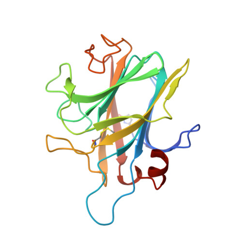
1D7B, 1D7C, 1D7D - PubMed Abstract:
The fungal oxidoreductase cellobiose dehydrogenase (CDH) degrades both lignin and cellulose, and is the only known extracellular flavocytochrome. This haemoflavoenzyme has a multidomain organisation with a b-type cytochrome domain linked to a large flavodehydrogenase domain. The two domains can be separated proteolytically to yield a functional cytochrome and a flavodehydrogenase. Here, we report the crystal structure of the cytochrome domain of CDH. The crystal structure of the b-type cytochrome domain of CDH from the wood-degrading fungus Phanerochaete chrysosporium has been determined at 1.9 A resolution using multiple isomorphous replacement including anomalous scattering information. Three models of the cytochrome have been refined: the in vitro prepared cytochrome in its redox-inactive state (pH 7.5) and redox-active state (pH 4.6), as well as the naturally occurring cytochrome fragment. The 190-residue long cytochrome domain of CDH folds as a beta sandwich with the topology of the antibody Fab V(H) domain. The haem iron is ligated by Met65 and His163, which confirms previous results from spectroscopic studies. This is only the second example of a b-type cytochrome with this ligation, the first being cytochrome b(562). The haem-propionate groups are surface exposed and, therefore, might play a role in the association between the cytochrome and flavoprotein domain, and in interdomain electron transfer. There are no large differences in overall structure of the cytochrome at redox-active pH as compared with the inactive form, which excludes the possibility that pH-dependent redox inactivation results from partial denaturation. From the electron-density map of the naturally occurring cytochrome, we conclude that it corresponds to the proteolytically prepared cytochrome domain.
- Department of Cell and Molecular Biology, Structural Biology, Uppsala University, Uppsala, 751 24, Sweden.
Organizational Affiliation: