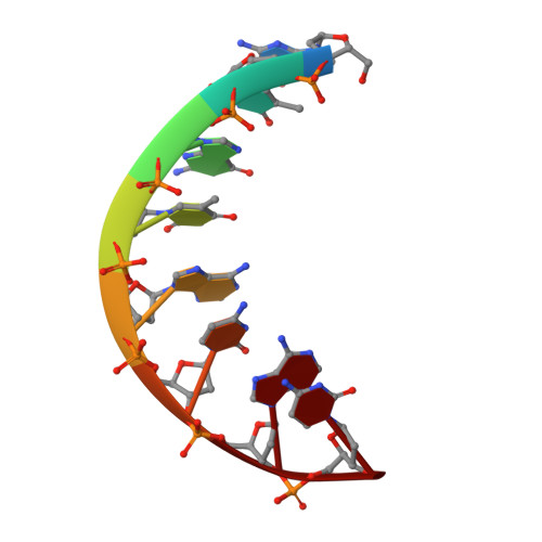
High-resolution refinement of the hexagonal A-DNA octamer d(GTGTACAC) at 1.4 A.
Thota, N., Li, X.H., Bingman, C.A., Sundaralingam, M.(1993) Acta Crystallogr D Biol Crystallogr 49: 282-291
- PubMed: 15299533 Search on PubMed
- DOI: https://doi.org/10.1107/S0907444992007522
- Primary Citation Related Structures:
1D78, 1D79 - PubMed Abstract:
The hexagonal crystal form of the octamer d(GTGTACAC), grown in the presence of spermine, has unit-cell dimensions a = b = 32.18 and c = 78.51 A, space group P6(1)22, with one DNA strand in the asymmetric unit. The structure has been refined starting with the earlier lower resolution model and using high-resolution 1.4 A data collected on a Siemens-Xentronics area detector at 258 K. There were 4365 unique reflections greater than 2sigma(F) in the resolution range 5-1.4 A. The model was refitted into 3F(o) - 2F(c). Sim-weighted omit maps and difference maps were used to locate water molecules. The final model with 161 DNA atoms and 37 water molecules gave an R factor of 19.8%. Crystals of the same octamer were also grown in the presence of spermidine instead of spermine, and refinement using nominal 1.45 A resolution data, 3292 unique reflections, final R = 19.1%, gave virtually identical DNA parameters. No bound spermine or spermidine was detected in either of these structure analyses. The electron density was clear for the DNA and showed holes in the center of the six-membered rings of bases, and also in the center of some of the sugar rings. The high-resolution structure has provided more precise DNA parameters and confirmed the features observed in the earlier 2 A study including the packing-induced distortion in the A7 (A15) sugar pucker from C(3')-endo and C(2')-endo. This change causes the end base pairs to bend away from the helix axis while the rest of the duplex is nearly linear. The hydration patterns in the deep and shallow grooves have been characterized. Chains of water molecules were found, but no rings. The familiar intermolecular contact region between the end base pair and the minor groove of a symmetry-related duplex, involving four residues on one strand and two on the other, has been analyzed. One of these interactions is a hydrogen bond.
- Crystallography Laboratory, Department of Biochemistry, College of Agricultural and Life Sciences, University of Wisconsin-Madison, 53706, USA.
Organizational Affiliation: