Effect of a single 3'-methylene phosphonate linkage on the conformation of an A-DNA octamer double helix.
Heinemann, U., Rudolph, L.N., Alings, C., Morr, M., Heikens, W., Frank, R., Blocker, H.(1991) Nucleic Acids Res 19: 427-433
- PubMed: 2011517 Search on PubMedSearch on PubMed Central
- DOI: https://doi.org/10.1093/nar/19.3.427
- Primary Citation Related Structures:
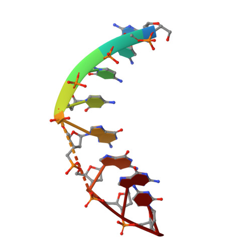
1D26 - PubMed Abstract:
The three-dimensional structure of the self-complementary DNA octamer d(GCCCGpGGC) has been determined in the crystalline state using X-ray diffraction data to a nominal resolutoin of 2.12 measured from a very small crystal at DESY, Hamburg. The structure was refined with stereochemical restraints to an R value of 17.1%. d(GCCCGpGGC), containing one single 3'-methylene phosphonate linkage (denoted p), forms an A-DNA double helix with strict dyad symmetry, that is distinct from canonical A-DNA by a wide open major groove and a small average base-pair inclination against the helix axis. The conformation of the unmodified control d(GCCCGGGC) is known from an X-ray analysis of isomorphous crystals (Heinemann et al. (1987) Nucleic Acids Res. 15, 9531-9550). Comparison of the two structures reveals only minor conformational differences, most notably in the pucker of the reduced deoxyribose. It is suggested that oligonucleotides with charged 3'-methylene phosphonate groups may form stable duplexes with complementary DNA or RNA strands rendering them candidates for use as gene-regulatory antisense probes.
- Abteilung Saenger, Freie Universität Berlin, FRG.
Organizational Affiliation: