Determination of the solution structure of a synthetic two-site calcium-binding homodimeric protein domain by NMR spectroscopy.
Shaw, G.S., Hodges, R.S., Sykes, B.D.(1992) Biochemistry 31: 9572-9580
- PubMed: 1390738 Search on PubMed
- DOI: https://doi.org/10.1021/bi00155a009
- Primary Citation Related Structures:
1CTA, 1CTD - PubMed Abstract:
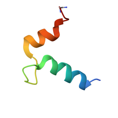
The solution structure of a 34-residue synthetic calcium-binding peptide from site III of chicken troponin-C has been determined by 1H NMR spectroscopy. In solution and in the presence of calcium this peptide forms a symmetric two-site homodimeric calcium-binding domain (Shaw et al., 1990). The solution structure of this dimer was determined from the measurement of 470 NOEs from a 75-ms NOESY data set. For the dimer structure determination, the constraint list included 868 distance restraints, 44 phi angles, and 24 chi 1 and 2 chi 2 angles. Seven structures were calculated by restrained molecular dynamics using a procedure in which intramonomer distances were used first and then all distances, intra- and intermonomer, were input during further dynamics. The structures exhibited a fold very similar to the C-terminal domain of troponin-C comprised of a pair of helix-loop-helix calcium-binding sites. The rms deviation of these structures for backbone atoms between residues 97-122 and 97'-122' for the dimer was 0.82 A. The dimer structure was also calculated to be more symmetric than sites III and IV in troponin-C.
- Department of Biochemistry, University of Alberta, Edmonton, Canada.
Organizational Affiliation: