Evolutionary constraints for dimer formation in prokaryotic Cu,Zn superoxide dismutase.
Bordo, D., Matak, D., Djinovic-Carugo, K., Rosano, C., Pesce, A., Bolognesi, M., Stroppolo, M.E., Falconi, M., Battistoni, A., Desideri, A.(1999) J Mol Biol 285: 283-296
- PubMed: 9878406 Search on PubMed
- DOI: https://doi.org/10.1006/jmbi.1998.2267
- Primary Citation Related Structures:
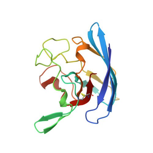
1BZO - PubMed Abstract:
Prokaryotic Cu,Zn superoxide dismutases are characterized by a distinct quaternary structure, as compared to that of the homologous eukaryotic enzymes. Here we report a newly determined crystal structure of the dimeric Cu,Zn superoxide dismutase from Photobacterium leiognathi (crystallized in space group R32, refined at 2.5 A resolution, R-factor 0.19) and analyse it in comparison with that of the monomeric enzyme from Escherichia coli. The dimeric assembly, observed also in a previously studied monoclinic crystal form of P. leiognathi Cu,Zn superoxide dismutase, is based on a ring-shaped subunit contact region, defining a solvated interface cavity. Three clusters of neighbouring residues play a direct role in the stabilization of the quaternary assembly. The present analysis, extended to the amino acid sequences of the other 11 known prokaryotic Cu,Zn superoxide dismutases, shows that at least in five other prokaryotic enzymes the interface residue clusters are under strong evolutionary constraint, suggesting the attainment of a quaternary structure coincident with that of P. leiognathi Cu,Zn superoxide dismutase. Calculation of electrostatic fields for both the enzymes from E. coli and P. leiognathi shows that the monomeric/dimeric association behaviour displayed by prokaryotic Cu, Zn superoxide dismutases is related to the distribution of surface charged residues. Moreover, Brownian dynamics simulations reproduce closely the observed enzyme:substrate association rates, highlighting the role of the active site neighbouring residues in determining the dismutase catalytic properties.
- Department of Physics - INFM and Advanced Biotechnology Center - IST, University of Genova, Largo R. Benzi, 10, Genova, I-16132, Italy.
Organizational Affiliation: