Crystal structure of the E2 DNA-binding domain from human papillomavirus type 16: implications for its DNA binding-site selection mechanism.
Hegde, R.S., Androphy, E.J.(1998) J Mol Biology 284: 1479-1489
- PubMed: 9878365 Search on PubMed
- DOI: https://doi.org/10.1006/jmbi.1998.2260
- Primary Citation Related Structures:
1BY9 - PubMed Abstract:
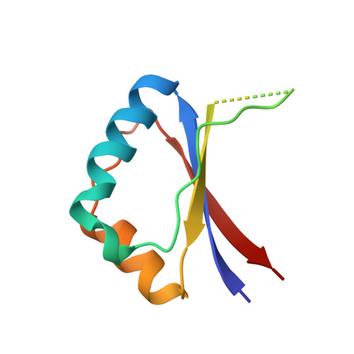
The crystal structure of the E2 DNA-binding domain from the high-risk cervical cancer-associated strain human papillomavirus type 16 (HPV-16) is described here. The papillomavirus E2 proteins regulate transcription from all viral promoters and are required for the initiation of replication in vivo. They belong to a family of viral proteins that form dimeric beta-barrels and use surface alpha-helices for DNA interaction. Although all E2 proteins recognize the same consensus, palindromic DNA sequence, proteins from different viral strains differ in their abilities to discriminate among their specific DNA-binding sites. The structure reported here reveals that while the overall fold of the HPV-16 E2 DNA-binding domain resembles that of its counterpart from the related viral strain bovine papillomavirus type 1, the precise placement of the recognition helices is significantly different. Additionally, the charge distribution on the DNA-binding surfaces of the two proteins varies; HPV-16 E2 has a much less electropositive surface. HPV-16 E2 is thus less able to utilize charge neutralization of the phosphate groups on DNA to induce bending. These results correlate well with previous solution studies that showed decreased affinity between HPV-16 E2 and flexible DNA target sequences, and enhanced affinity towards A-tract-containing, pre-bent sequences. In summary, the crystal structure of the HPV-16 E2 DNA-binding domain shows that the protein presents a stereo-chemically and electrostatically unique surface to DNA, characteristics that can contribute to its mechanism of DNA target discrimination.
- Department of Biochemistry and Program in Structural Biology, New York University Medical Center, Skirball Institute of Biomolecular Medicine, 540 First Avenue, New York, NY, 10016, USA. rashmi@saturn.med.nyu.edu
Organizational Affiliation: