1BXX

MU2 ADAPTIN SUBUNIT (AP50) OF AP2 ADAPTOR (SECOND DOMAIN), COMPLEXED WITH TGN38 INTERNALIZATION PEPTIDE DYQRLN


Experimental Data Snapshot

  • Method: X-RAY DIFFRACTION
  • Resolution: 2.70 Å
  • R-Value Free: 0.325 
  • R-Value Work: 0.263 
  • R-Value Observed: 0.282 

wwPDB Validation   3D Report Full Report


This is version 1.4 of the entry. See complete history


Literature

A structural explanation for the recognition of tyrosine-based endocytotic signals.

Owen, D.J.Evans, P.R.

(1998) Science 282: 1327-1332

  • DOI: https://doi.org/10.1126/science.282.5392.1327
  • Primary Citation of Related Structures:  
    1BW8, 1BXX

  • PubMed Abstract: 

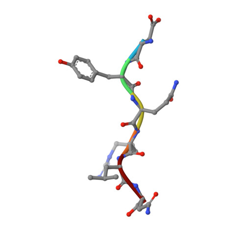
    Many cell surface proteins are marked for endocytosis by a cytoplasmic sequence motif, tyrosine-X-X-(hydrophobic residue), that is recognized by the mu2 subunit of AP2 adaptors. Crystal structures of the internalization signal binding domain of mu2 complexed with the internalization signal peptides of epidermal growth factor receptor and the trans-Golgi network protein TGN38 have been determined at 2.7 angstrom resolution. The signal peptides adopted an extended conformation rather than the expected tight turn. Specificity was conferred by hydrophobic pockets that bind the tyrosine and leucine in the peptide. In the crystal, the protein forms dimers that could increase the strength and specificity of binding to dimeric receptors.


  • Organizational Affiliation

    Medical Research Council Laboratory of Molecular Biology, Hills Road, Cambridge CB2 2QH, UK.


Macromolecules
Find similar proteins by:  (by identity cutoff)  |  3D Structure
Entity ID: 1
MoleculeChains Sequence LengthOrganismDetailsImage
PROTEIN (AP50)285Rattus norvegicusMutation(s): 0 
UniProt
Find proteins for P84092 (Rattus norvegicus)
Explore P84092 
Go to UniProtKB:  P84092
Entity Groups  
Sequence Clusters30% Identity50% Identity70% Identity90% Identity95% Identity100% Identity
UniProt GroupP84092
Sequence Annotations
Expand
  • Reference Sequence

Find similar proteins by:  Sequence   |   3D Structure  

Entity ID: 2
MoleculeChains Sequence LengthOrganismDetailsImage
PROTEIN (TGN38 PEPTIDE)B [auth P]6synthetic constructMutation(s): 0 
Sequence Annotations
Expand
  • Reference Sequence
Experimental Data & Validation

Experimental Data

  • Method: X-RAY DIFFRACTION
  • Resolution: 2.70 Å
  • R-Value Free: 0.325 
  • R-Value Work: 0.263 
  • R-Value Observed: 0.282 
  • Space Group: P 64
Unit Cell:
Length ( Å )Angle ( ˚ )
a = 125.26α = 90
b = 125.26β = 90
c = 73.79γ = 120
Software Package:
Software NamePurpose
REFMACrefinement
MOSFLMdata reduction
CCP4data scaling

Structure Validation

View Full Validation Report


Entry History 

Deposition Data

Revision History  (Full details and data files)

  • Version 1.0: 1998-11-25
    Type: Initial release
  • Version 1.1: 2008-04-27
    Changes: Version format compliance
  • Version 1.2: 2011-07-13
    Changes: Version format compliance
  • Version 1.3: 2018-05-30
    Changes: Data collection, Source and taxonomy, Structure summary
  • Version 1.4: 2023-08-09
    Changes: Data collection, Database references, Refinement description