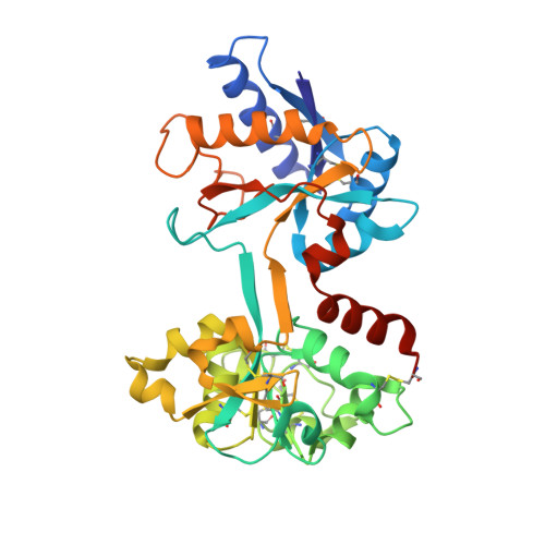
Ligand-induced conformational change in transferrins: crystal structure of the open form of the N-terminal half-molecule of human transferrin.
Jeffrey, P.D., Bewley, M.C., MacGillivray, R.T., Mason, A.B., Woodworth, R.C., Baker, E.N.(1998) Biochemistry 37: 13978-13986
- PubMed: 9760232 Search on PubMed
- DOI: https://doi.org/10.1021/bi9812064
- Primary Citation Related Structures:
1BP5, 1BTJ - PubMed Abstract:
Serum transferrin binds ferric ions in the bloodstream and transports them to cells, where they are released in a process involving receptor-mediated endocytosis. Iron release is believed to be pH dependent and is coupled with a large conformational change. To help define the steps in iron release, we have determined the three-dimensional structure of the iron-free (apo) form of the recombinant N-lobe half-molecule of human serum transferrin (ApoTfN) by X-ray crystallography. Two crystal forms were obtained, form 1 with four molecules in the asymmetric unit and form 2 with two molecules in the asymmetric unit. The structures of both forms were determined by molecular replacement and were refined at 2.2 and 3.2 A resolution, respectively. Final R-factors were 0.203 (free R = 0. 292) for form 1 and 0.217 (free R = 0.312) for form 2. All six copies of the ApoTfN structure are essentially identical. Comparison with the holo form (FeTfN) shows that a large rigid-body domain movement of 63 degrees has occurred in ApoTfN, to give an open binding cleft. The extent of domain opening is the same as in the N-lobe of human lactoferrin, showing that it depends on internal constraints that are conserved in both proteins, and that it is unaffected by the presence or absence of the C-lobe. Although the conformational change is primarily a rigid-body motion, several local adjustments occur. In particular, two iron ligands, Asp 63 and His 249, change conformation to form salt bridges, with Lys 296 and Glu 83, respectively, in the binding cleft of the apo protein. Both salt bridges would have to break for iron coordination to occur. Most importantly, the structure, determined at a pH (5.3) that is close to the pH of physiological iron release, indicates that protonation of His 249 is a key step in iron release.
- Department of Biochemistry, Massey University, Palmerston North, New Zealand.
Organizational Affiliation: