1BHX

X-RAY STRUCTURE OF THE COMPLEX OF HUMAN ALPHA THROMBIN WITH THE INHIBITOR SDZ 229-357


Experimental Data Snapshot

  • Method: X-RAY DIFFRACTION
  • Resolution: 2.30 Å
  • R-Value Free: 0.232 
  • R-Value Work: 0.187 
  • R-Value Observed: 0.187 

wwPDB Validation   3D Report Full Report


Ligand Structure Quality Assessment 


This is version 1.5 of the entry. See complete history


Literature

Rational design, synthesis, and X-ray structure of selective noncovalent thrombin inhibitors.

Wagner, J.Kallen, J.Ehrhardt, C.Evenou, J.P.Wagner, D.

(1998) J Med Chem 41: 3664-3674

  • DOI: https://doi.org/10.1021/jm981013e
  • Primary Citation of Related Structures:  
    1BHX

  • PubMed Abstract: 

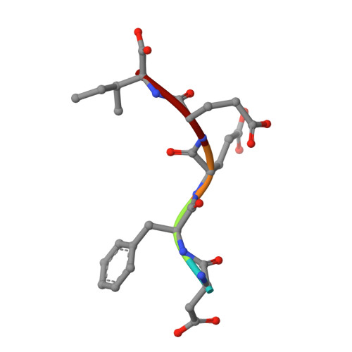
    We have designed, synthesized, and tested in vitro a novel class of noncovalent thrombin inhibitors. The main feature of these inhibitors is a 6,5-fused bicyclic core structure that fills the S2 pocket of the active site of thrombin. The bicycle introduces conformational constraint into the ligand and locks the Xaa-Pro amide bond into the desired trans configuration. Among the known ring systems, we selected by molecular modeling the 7-thiaindolizidinones (BTD) as our basic template. The influence of several structural features was analyzed: the length of the argininal side chain, the stereochemistry at C6, and the importance of making optimal use of the S3 pocket. Finally, an X-ray crystal structure of inhibitor 15 bound to thrombin was obtained at a resolution of 2.3 A. These designed thrombin inhibitors, which were prepared by an efficient synthesis, showed high selectivity over trypsin and other serine proteases. Further derivation based on the information obtained by X-ray crystallography should certainly allow to improve the potency.


  • Organizational Affiliation

    Novartis Pharma AG, CH-4002 Basel, Switzerland.


Macromolecules
Find similar proteins by:  (by identity cutoff)  |  3D Structure
Entity ID: 1
MoleculeChains Sequence LengthOrganismDetailsImage
ALPHA THROMBIN30Homo sapiensMutation(s): 0 
EC: 3.4.21.5
UniProt & NIH Common Fund Data Resources
Find proteins for P00734 (Homo sapiens)
Explore P00734 
Go to UniProtKB:  P00734
PHAROS:  P00734
GTEx:  ENSG00000180210 
Entity Groups  
Sequence Clusters30% Identity50% Identity70% Identity90% Identity95% Identity100% Identity
UniProt GroupP00734
Sequence Annotations
Expand
  • Reference Sequence
Find similar proteins by:  (by identity cutoff)  |  3D Structure
Entity ID: 2
MoleculeChains Sequence LengthOrganismDetailsImage
ALPHA THROMBIN147Homo sapiensMutation(s): 0 
EC: 3.4.21.5
UniProt & NIH Common Fund Data Resources
Find proteins for P00734 (Homo sapiens)
Explore P00734 
Go to UniProtKB:  P00734
PHAROS:  P00734
GTEx:  ENSG00000180210 
Entity Groups  
Sequence Clusters30% Identity50% Identity70% Identity90% Identity95% Identity100% Identity
UniProt GroupP00734
Sequence Annotations
Expand
  • Reference Sequence
Find similar proteins by:  (by identity cutoff)  |  3D Structure
Entity ID: 3
MoleculeChains Sequence LengthOrganismDetailsImage
ALPHA THROMBINC [auth F]105Homo sapiensMutation(s): 0 
EC: 3.4.21.5
UniProt & NIH Common Fund Data Resources
Find proteins for P00734 (Homo sapiens)
Explore P00734 
Go to UniProtKB:  P00734
PHAROS:  P00734
GTEx:  ENSG00000180210 
Entity Groups  
Sequence Clusters30% Identity50% Identity70% Identity90% Identity95% Identity100% Identity
UniProt GroupP00734
Sequence Annotations
Expand
  • Reference Sequence

Find similar proteins by:  Sequence   |   3D Structure  

Entity ID: 4
MoleculeChains Sequence LengthOrganismDetailsImage
ALPHA THROMBIND [auth E]5Homo sapiensMutation(s): 0 
EC: 3.4.21.5
Sequence Annotations
Expand
  • Reference Sequence
Small Molecules
Ligands 1 Unique
IDChains Name / Formula / InChI Key2D Diagram3D Interactions
R56
Query on R56

Download Ideal Coordinates CCD File 
E [auth F]5-OXO-6-PHENYLMETHANESULFONYLAMINO-HEXAHYDRO-THIAZOLO[3,2-A]PYRIDINE-3-CARBOXYLIC ACID (3-GUANIDINO-PROPYL)-AMIDE
C19 H28 N6 O4 S2
SOIIHESTBYNJRH-PMPSAXMXSA-N
Binding Affinity Annotations 
IDSourceBinding Affinity
R56 Binding MOAD:  1BHX Ki: 145 (nM) from 1 assay(s)
PDBBind:  1BHX Ki: 145 (nM) from 1 assay(s)
Experimental Data & Validation

Experimental Data

  • Method: X-RAY DIFFRACTION
  • Resolution: 2.30 Å
  • R-Value Free: 0.232 
  • R-Value Work: 0.187 
  • R-Value Observed: 0.187 
  • Space Group: C 1 2 1
Unit Cell:
Length ( Å )Angle ( ˚ )
a = 71α = 90
b = 72.2β = 100.7
c = 73.1γ = 90
Software Package:
Software NamePurpose
MADNESdata collection
PROCORdata reduction
Agrovatadata reduction
X-PLORmodel building
X-PLORrefinement
MADNESdata reduction
PROCORdata scaling
Agrovatadata scaling
X-PLORphasing

Structure Validation

View Full Validation Report


Ligand Structure Quality Assessment 


Entry History 

Deposition Data

  • Released Date: 1998-11-04 
  • Deposition Author(s): Kallen, J.

Revision History  (Full details and data files)

  • Version 1.0: 1998-11-04
    Type: Initial release
  • Version 1.1: 2008-03-24
    Changes: Version format compliance
  • Version 1.2: 2011-07-13
    Changes: Version format compliance
  • Version 1.3: 2011-11-16
    Changes: Atomic model
  • Version 1.4: 2018-04-18
    Changes: Data collection, Other, Refinement description
  • Version 1.5: 2023-08-02
    Changes: Database references, Derived calculations, Refinement description